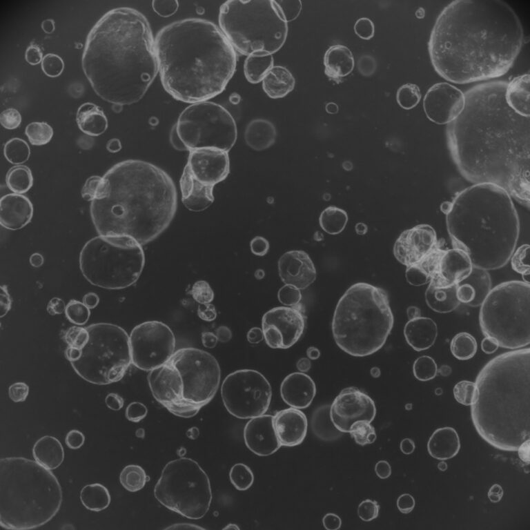
Alzheimer’s disease is often discussed as a single disorder, yet patients can experience remarkably different symptoms and respond differently to the same treatment. A recent study from Johns Hopkins Medicine adds to growing evidence that patient-derived brain organoids—miniature brain tissues grown from an individual’s own cells—may help explain these differences and support more personalized therapeutic strategies.
Using induced pluripotent stem cells (iPSCs) generated from the blood of patients with Alzheimer’s disease, researchers created hundreds of hindbrain organoids containing serotonin-producing neurons. Proteomic analysis showed that these organoids faithfully reproduced molecular features associated with Alzheimer’s disease, including disruptions in neuronal communication, inflammatory pathways, and proteins involved in synaptic signaling.
The team then examined how these organoids responded to escitalopram, a selective serotonin reuptake inhibitor (SSRI) commonly prescribed to manage neuropsychiatric symptoms such as anxiety, depression, and agitation in Alzheimer’s patients. While some organoids exhibited increased expression of proteins linked to serotonin signaling and synaptic function following treatment, others showed little or no response. This heterogeneity suggests that patients may differ substantially at the molecular level, even when receiving the same medication.
An equally significant finding involved extracellular vesicles (EVs) released by the organoids. These microscopic particles carried protein signatures reflecting disease state and treatment response. Several proteins involved in neuronal signaling—including RAB3A, NSF, and ATCAY—were reduced in Alzheimer’s-derived organoids, while escitalopram altered vesicle protein profiles only in responsive samples. The authors propose that EVs could eventually serve as minimally invasive biomarkers for disease staging and treatment stratification.
Although still at an early stage, this work demonstrates the potential of combining brain organoid technology with large-scale proteomic profiling to capture patient-specific biology. Rather than viewing Alzheimer’s as a uniform disease, these findings support a future where therapies can be matched to molecular subtypes, improving treatment selection while accelerating biomarker discovery. As brain organoid models continue to incorporate additional physiological features such as immune cells and vascular networks, they may become an increasingly valuable platform for precision neuroscience research.
Research Article: Boyd RJ, Dong D, Sagar R, et al. Proteomic profiling of brain organoids and extracellular vesicles identifies early Alzheimer’s disease biomarkers and drug response heterogeneity. Alzheimer’s Dement. 2026; 22:e71273. https://doi.org/10.1002/alz.71273
Working on similar research topics?
Explore our brain organoid platform and Neurodegeneration disease models