
Retinal organoids are transforming the way researchers study human retinal development, inherited retinal diseases, and therapeutic responses. Generated from stem cells, these three-dimensional retinal models can reproduce key cellular and structural features of the human retina, making them valuable tools for disease modeling, drug screening, and regenerative medicine research.
Based on insights from the review “Retinal Organoids: Advances in Generation, Development, and Applications – From Stem Cells to Disease Modeling and Regenerative Medicine”, this blog explores how retinal organoids are advancing ophthalmology research and introduces Lambda’s retinal organoid platform for human-relevant retinal disease modeling and therapeutic development.
In This Article
What Are Retinal Organoids?
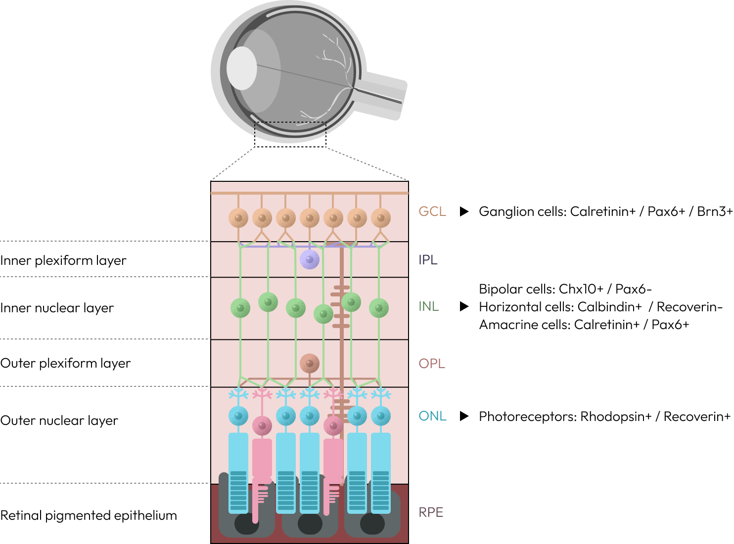
Retinal organoids are three-dimensional, stem cell-derived tissue models that recapitulate key features of the human retina. They can be generated from embryonic stem cells or induced pluripotent stem cells, also known as iPSCs, and are able to self-organize into retinal-like structures under defined culture conditions.
Unlike traditional two-dimensional cell cultures, retinal organoids contain multiple retinal cell populations arranged in a more tissue-like architecture. These may include retinal progenitor cells, photoreceptors, retinal ganglion cells, interneurons, Müller glia, and retinal pigment epithelium-like cells, depending on the protocol and maturation stage.
Because of their human origin and three-dimensional organization, retinal organoids are increasingly used as advanced in vitro models for studying retinal development, retinal disease mechanisms, therapeutic responses, and regenerative medicine strategies.
Why Retinal Organoids Matter in Eye Disease Research
Many retinal diseases are difficult to study using conventional models. Animal models have contributed significantly to ophthalmology research, but they cannot fully reproduce all aspects of the human eye. For example, the human retina has features such as the macula and complex color vision that are not accurately represented in many animal systems.
Retinal organoids help address this limitation by providing a human-relevant model system. When derived from patient-specific iPSCs, they can retain the genetic background of individuals with retinal disease. This allows researchers to study how specific mutations affect retinal cell development, tissue organization, photoreceptor maturation, ciliary function, synaptic development, and cell survival.
As a result, retinal organoids are becoming an important bridge between basic research, preclinical development, and precision medicine.
Retinal Organoids for Inherited Retinal Disease Modeling
One of the most important applications of retinal organoids is the study of inherited retinal diseases, or IRDs. IRDs are a diverse group of genetic disorders that cause progressive vision loss due to dysfunction or degeneration of retinal cells, especially photoreceptors and retinal pigment epithelial cells.
IRDs may appear in childhood, adolescence, or adulthood. They may affect only the retina, or they may occur as part of broader syndromic diseases involving other organs. Their genetic complexity makes them challenging to diagnose and treat, with hundreds of genes implicated in different forms of retinal degeneration.
Patient-derived iPSC retinal organoids offer a powerful way to model these diseases in vitro. By generating retinal tissue from patients carrying disease-associated mutations, researchers can observe how those mutations alter cellular development and retinal function.
Examples of retinal diseases that can be modeled using retinal organoids include:
- Leber congenital amaurosis, including disease associated with ciliary genes such as CEP290
- Retinitis pigmentosa, including forms linked to genes such as USH2A
- Stargardt disease, commonly associated with ABCA4 mutations
- Glaucoma-related retinal ganglion cell degeneration
- Microphthalmia, a developmental eye disorder
- Retinoblastoma, a childhood retinal cancer associated with RB1 inactivation
These models allow researchers to connect genotype with phenotype. In practical terms, this means scientists can study how a specific genetic alteration leads to measurable disease features such as defective ciliogenesis, impaired photoreceptor development, endoplasmic reticulum stress, apoptosis, or abnormal retinal cell organization.
Retinal Organoids in Drug Screening, Development, and Regenerative Medicine
Beyond disease modeling, retinal organoids are increasingly used for drug screening and therapeutic discovery. Their three-dimensional, multicellular structure provides a more physiologically relevant system than conventional 2D cultures, making them useful for evaluating small molecules, gene therapies, RNA-based approaches, neuroprotective compounds, toxicity signals, and disease-specific biomarker responses.
Retinal organoids also support precision medicine, particularly for inherited retinal diseases with high genetic diversity. Patient-specific organoids can help researchers study how different mutations influence disease progression and treatment response.
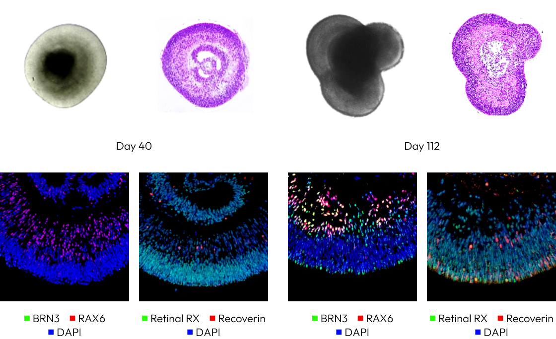
In developmental biology, retinal organoids provide a controllable model for studying human retinal formation, including retinal progenitor specification, neuronal differentiation, photoreceptor maturation, and optic vesicle-like development. This is especially valuable because access to human developmental retinal tissue is limited.
For regenerative medicine, retinal organoid-derived cells, such as retinal progenitor cells and photoreceptor precursors, are being explored as potential sources for retinal cell replacement. However, key challenges remain, including cell purity, long-term safety, tumorigenic risk, functional integration, and synaptic connectivity with host retinal tissue.
Despite their promise, retinal organoids still face limitations such as batch-to-batch variability, incomplete maturation, limited light responsiveness, and a lack of full interaction with supporting components like mature RPE, vasculature, immune cells, and microglia. Advanced co-culture systems, retinal organoid-RPE models, and organ-on-a-chip technologies are being developed to improve maturity, reproducibility, and translational relevance.
Introducing Lambda’s Retinal Organoid Platform
Lambda’s retinal organoid platform is designed to support human-relevant retinal research, disease modeling, and therapeutic development. By using stem cell-derived three-dimensional retinal tissue models, Lambda provides researchers with an advanced in vitro system that more closely reflects the complexity of human retinal biology than conventional flat cell culture systems.
The platform can support research across multiple areas of retinal biology, including retinal progenitor differentiation, photoreceptor maturation, retinal ganglion cell biology, ciliogenesis-associated defects, degeneration-related stress responses, and disease-relevant biomarker analysis.
Lambda’s retinal organoid platform is particularly relevant for studies involving inherited retinal diseases, photoreceptor degeneration, Leber congenital amaurosis, retinitis pigmentosa, Stargardt disease, glaucoma-related retinal ganglion cell damage, and retinal safety assessment.
Applications:
- Disease Modeling
- Drug Discovery and Screening
- Gene and RNA Therapy Assessment
- Biomarker and Phenotypic Analysis
- Translational Ophthalmology Research


