Fibrosis has traditionally been studied one organ at a time — pulmonologists track idiopathic pulmonary fibrosis (IPF), gastroenterologists track fibrostenotic Crohn’s disease, hepatologists track liver cirrhosis — each field running its own animal models, biomarkers, and drug pipelines. But a growing body of research treats fibrosis as a single disease process with a shared molecular signature, regardless of which organ is scarring: sustained TGF-β/Smad-driven myofibroblast activation and excessive extracellular matrix (ECM) deposition. Some researchers describe fibrosis as a “final common pathway” toward organ failure that happens to present differently depending on where it occurs — a framing first articulated by Border and Noble in their work on angiotensin II and tissue repair, and reiterated in Rockey, Bell, and Hill’s 2015 review in the New England Journal of Medicine.
Lambda Biologics has developed two human organoid–based fibrosis models — one built on human lung organoids, the other on human intestinal organoids — that capture this shared biology in 3D, tissue-relevant systems. Looking at them together clarifies a point that matters for antifibrotic drug discovery: a compound’s mechanism of action doesn’t necessarily stay confined to the organ it was designed for.
In This Article
Fibrosis Is a Convergent Process, Not an Organ-Specific One
Across the lung, gut, liver, kidney, and heart, fibrogenesis converges on the same core cascade: chronic injury activates fibroblasts and related effector cells, which differentiate into myofibroblasts under TGF-β/Smad2/3 signaling and begin depositing collagen and other ECM proteins faster than the tissue can clear them. Reviews of the TGF-β/Smad pathway in fibrosis describe this axis as central to fibrogenesis in essentially every solid organ, and a 2025 review calling for a unified, multi-organ approach to antifibrotic drug development points to conserved regulatory nodes – including PPAR signaling, NOX4/Nrf2 oxidative stress pathways, and specific HDAC isoforms – that recur across liver, lung, kidney, and cardiac fibrosis alike. This shared mechanism already shows up in the clinic. Nintedanib, approved for IPF, has documented antifibrotic activity in preclinical models of the liver, skin, and kidney, and both nintedanib and pirfenidone are now used in interstitial lung diseases well beyond their original IPF indication. If a drug’s antifibrotic activity is mechanism-driven rather than organ-specific, then a model system capable of testing that mechanism in more than one tissue becomes considerably more useful.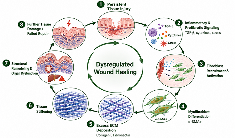
The Gut-Lung Connection Isn’t Just a Metaphor
Beyond the shared molecular pathway, there’s a more literal connection between pulmonary and intestinal fibrosis. Emerging research on the gut-lung axis in pulmonary fibrosis shows that IPF patients frequently present with intestinal dysbiosis and altered gut permeability, linked to circulating immune cell phenotypes via shared mucosal immune signaling. In parallel, studies of Crohn’s disease-associated fibrosis have identified neutrophil-fibroblast crosstalk as a driver of “immunofibrosis” in the gut – a mechanism with clear parallels to epithelial-stromal crosstalk defects observed in IPF lung tissue.
This is still an active, hypothesis-generating area of research rather than settled biology – the gut-lung axis literature is stronger on microbiome and immune signaling than on direct fibrotic crosstalk. But it reinforces the case for studying lung and intestinal fibrosis side by side rather than in isolation: what’s learned about epithelial-stromal dysfunction in one organoid platform may well be informative for the other.
Modeling Pulmonary Fibrosis in a Human Lung Organoid
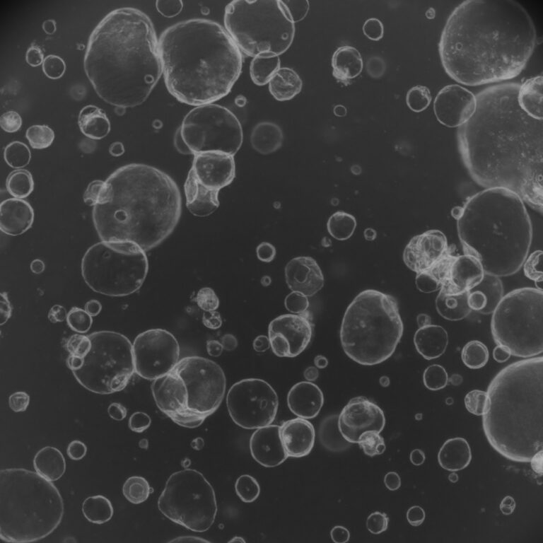
Lambda Biologics’ lung organoid platform is generated from human lung epithelial progenitor cells – either tissue-derived or iPSC-derived – and self-organizes into distal lung architecture containing type I and type II alveolar epithelial cells, ciliated cells, and secretory cells (confirmed via markers including SFTPC, AGER, AQP5, and FOXJ1). Fibrosis is induced through pro-fibrotic stimulation, most commonly TGF-β, alongside optional mechanical stress or fibroblast co-culture, producing fibroblast activation, excessive collagen I and fibronectin deposition, and measurable tissue stiffening – hallmarks of early-stage IPF.
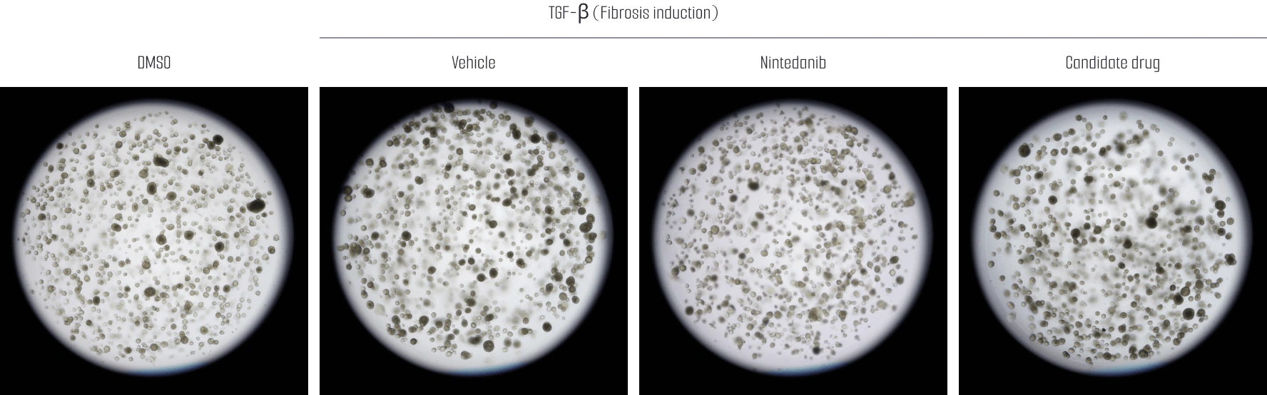
The model has been validated against nintedanib: treatment significantly reduced α-SMA and vimentin expression, confirming that the platform responds appropriately to a known clinical therapeutic. Several candidate compounds produced comparable, and in some cases statistically significant, reductions in fibrosis markers, supporting the model’s use for comparative antifibrotic screening. Full workflow and data are available in the pulmonary fibrosis case study.
Modeling Intestinal Fibrosis in a Human Intestinal Organoid
Intestinal fibrosis is a major complication of chronic inflammatory bowel disease, particularly Crohn’s disease, where progressive ECM deposition leads to luminal narrowing and stricture. Anti-inflammatory biologics have improved disease control, but effective antifibrotic therapies remain an unmet need, partly because inflammation-focused models don’t capture fibrotic remodeling on its own.
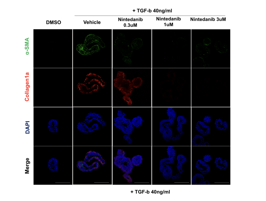
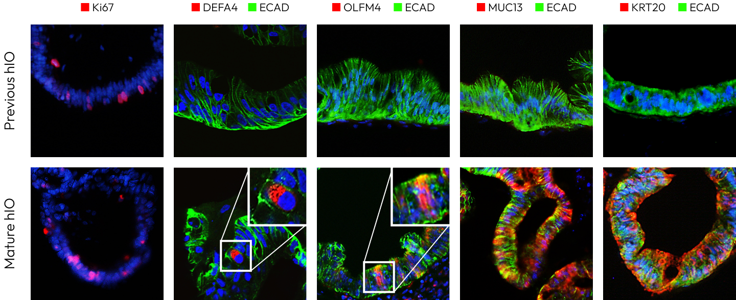
Lambda Biologics’ intestinal organoid fibrosis model addresses this by integrating epithelial organoids with stromal components, reconstructing the multicellular interactions that drive fibrogenesis in the human gut. The platform reproduces sustained fibroblast activation and myofibroblast differentiation, excessive collagen and ECM protein deposition, measurable tissue stiffening and architectural distortion, and activation of canonical TGF-β-associated signaling. Readouts include immunofluorescence quantification of α-SMA and collagen I/III, transcriptomic profiling of fibrosis-related genes, ECM and matrix composition analysis, functional stiffness assessment, and drug-response profiling. Full details are in the intestinal fibrosis case study; a related leaky gut syndrome model covers barrier dysfunction specifically, for programs that need to separate inflammation from fibrotic remodeling.
The Model’s Impacts
Running lung and intestinal organoid fibrosis models side by side, using a consistent readout set (α-SMA, collagen I/III, ECM composition, transcriptomics), lets researchers ask a question that single-organ models can’t answer on their own: is a candidate’s antifibrotic activity tied to lung-specific biology, or does it act on the conserved TGF-β/myofibroblast axis broadly enough to be relevant across indications? That question matters for indication expansion strategy, basket-trial design, and early go/no-go decisions – and it’s far cheaper to answer with two organoid platforms than with two animal studies.
Both platforms are screening-ready and reproducible, reducing reliance on animal models in line with New Approach Methodology (NAM) principles, and sit alongside Lambda Biologics’ broader disease modeling portfolio spanning liver, skin, brain, and additional organ systems.
Frequently Asked Questions
Are pulmonary fibrosis and intestinal fibrosis related?
Can the same antifibrotic drug work in both the lung and the gut?
What is an organoid fibrosis model?
Why use organoids instead of animal models for fibrosis research?
Lambda Biologics’s Organoid-based Discovery platform for Innovative Screening, Evaluation, and Identification