Beyond Plaques and Tangles
Alzheimer’s disease is often defined by amyloid-β plaques and tau tangles. Yet new research suggests that understanding the disease also requires knowing where cells are located, which neighbours surround them and how those relationships shape their behaviour.
Using a brain-adapted spatial-proteomics workflow called CODEX-CNS, Paula Sanchez-Molina and her team of researchers profiled 704,706 cells from the frontal cortex of eight people with Alzheimer’s disease and eight healthy controls. Unlike methods that require tissue dissociation, the approach preserved tissue architecture while mapping proteins, cell morphology, blood vessels, glia and amyloid pathology within the same sections.
Identity Shaped by Place
The analysis identified a disease-enriched population termed human plaque-associated microglia, or HPAM. These cells expressed markers associated with phagocytosis, antigen presentation and macrophage-like activity, and were closely associated with dense rather than diffuse amyloid plaques.
Importantly, the surrounding cellular neighbourhood predicted dense-plaque association more effectively than microglial morphology alone. This shifts attention from isolated cell types toward Alzheimer’s disease as a changing cellular ecosystem involving microglia, reactive astrocytes, vasculature, amyloid deposits and damaged neurites.
The finding does not yet establish whether HPAM protects neurons, intensifies inflammation or performs different roles as pathology progresses. Because the study examined postmortem tissue, it provides a detailed spatial snapshot rather than a record of how these states emerge over time.
From Spatial Maps to Testable Mechanisms
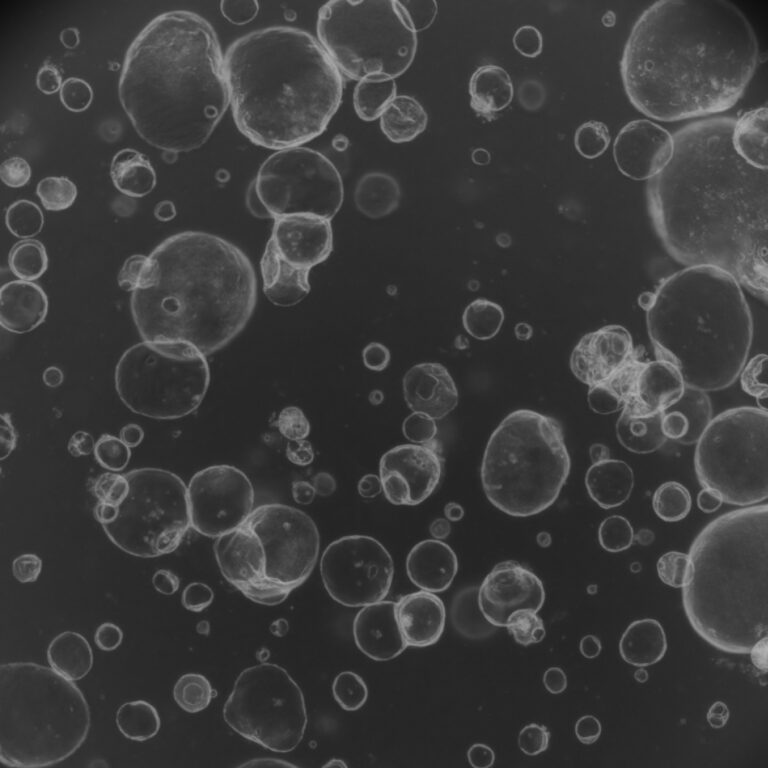
As Alzheimer’s research moves toward increasingly complex questions about multicellular interactions, iPSC-derived brain organoids offer a complementary way to begin investigating them. These three-dimensional human models allow researchers to vary genetic backgrounds, pathological stimuli and treatment conditions while observing cellular responses over time.
Advanced systems can also incorporate iPSC-derived microglia. In one long-term brain-organoid study, integrated microglia were associated with greater synaptic density and reduced phosphorylated tau, cellular debris and neuronal death—demonstrating how adding immune components can substantially alter model behaviour.
Organoids cannot reproduce the full ageing, vascular or peripheral immune environment of an adult brain. Their value is not in recreating Alzheimer’s disease in its entirety, but in helping convert spatial observations from human tissue into experimentally testable questions. Together, spatial proteomics and human organoid models could connect detailed disease maps with faster mechanistic studies and more human-relevant evidence for later translational validation.
Research article: Spatial proteomic analysis in human Alzheimer’s disease brains enables identification of microenvironment-dependent microglial cell states.
References
- Sanchez-Molina, P., Rosmus, D.-D., Brownell, D., et al. “Spatial proteomic analysis in human Alzheimer’s disease brains enables identification of microenvironment-dependent microglial cell states.” Nature Neuroscience 29, 1599–1614 (2026). DOI: 10.1038/s41593-026-02267-3.
- Chen, X., Sun, G., Feng, L., et al. “Human iPSC-derived microglial cells protect neurons from neurodegeneration in long-term cultured adhesion brain organoids.” Communications Biology 8, 30 (2025). DOI: 10.1038/s42003-024-07401-0.
Working on Brain research?
Explore our brain organoid platform and Neurodegeneration disease models