
Home » AFS » Striatal Organoid
Striatal-Organoids-main
Organoid Service

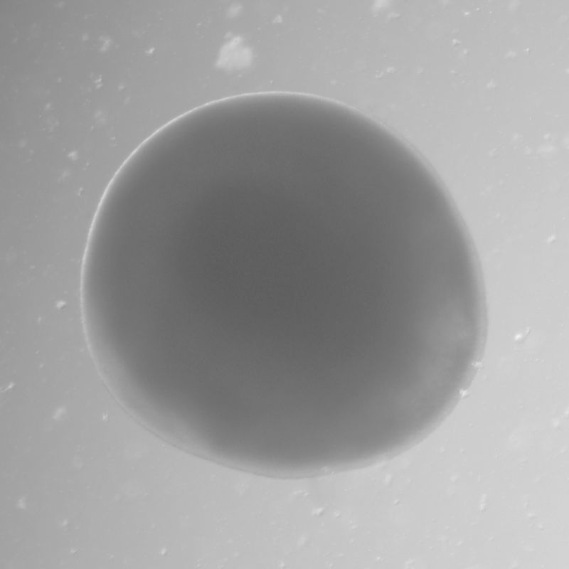
Striatal Organoid

Striatal Organoid

  • Human Medium Spiny Neuron Model
    Striatal organoids are 3D human brain organoids derived from induced pluripotent stem cells (iPSCs) that generate medium spiny neurons (MSNs), the principal neurons of the striatum. They give researchers access to human striatal neurons that are otherwise difficult to study directly.
  • Drug Efficacy & Neurotoxicity Testing
    Striatal organoids provide a human-relevant platform for evaluating neurological drug efficacy, neurotoxicity, and neuronal function, helping de-risk CNS drug discovery before clinical studies.
  • Movement Disorder Disease Modeling
    Striatal organoids can be genetically modified to investigate the mechanisms underlying Huntington's disease, Parkinson's disease, and other movement disorders.

Price
Organism
Human
Product Type
iPS-derived organoid
Tissue
Brain (striatal)
Disease
Neurodegenerative diseases, Brain tumor, Neurodevelopmental disorders

Applications

Toxicity

Organoid Based

Professor Lee Chang-seok Eulji University
Customer insight

Advancing K-Beauty with Skin Organoids

Advancing K-Beauty with Skin Organoids: A Next-Generation Platform for Non-Animal Testing and High-Precision Cosmetic Innovation With the global rise of K-beauty, the cosmetics industry continues to grow steadily. Since the...

Tomocube (Spatial)
Customer insight

HT-X1: A Label-Free Imaging Breakthrough for Organoid-Based Disease Modeling and Drug Screening

Traditional microscopy methods often require fluorescent labeling to analyze cellular structures, which can be time-consuming and invasive. In contrast, our HT-X1 system allows for high-resolution visualization of cellular morphology without...

Seoul National University College of Medicine
Customer insight

Advancing Clinical Pathology with Spatial Protein Analysis

Traditional protein analysis has primarily focused on quantifying expression levels within tissue samples. However, recent advances in spatial analysis techniques have shifted attention toward evaluating not only expression levels, but...

K Research Institute
Customer insight

Revealing Immune-Boosting Potential with ODISEI-Gut

Among the many fermented foods we consume, kimchi is particularly known for containing a diverse range of lactic acid bacteria, which are believed to influence the activation of immune cells...

Bundang Jesaeng General Hospital
Customer insight

Multiplex Marker Analysis Enhances Research Efficiency with 31-Marker Detection on a Single Slide

We conducted a study focused on identifying disease-related markers using patient-derived tissue samples. However, traditional methods limited our ability to analyze multiple candidate markers simultaneously, and the limited availability of...

Description

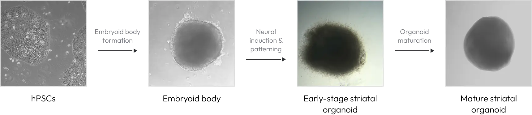
Striatal Organoid Differentiation Timeline

Our striatal organoid differentiation protocol has been validated across multiple human pluripotent stem cell (hPSC) lines, consistently generating striatal organoids that express key striatal markers, including DARPP32 and CTIP2, and exhibit the characteristics of medium spiny neurons (MSNs). From embryoid body formation to striatal maturation, each stage is guided by defined patterning cues that mirror human forebrain development.
Striatal organoid development from hPSCs

Medium Spiny Neuron Population in Striatal Organoids

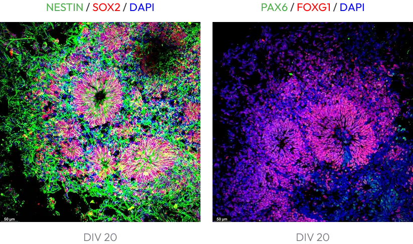
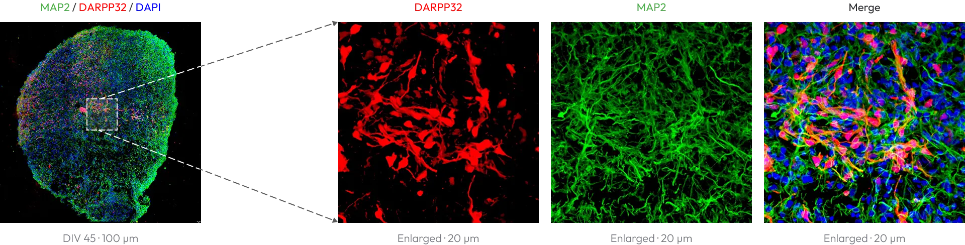
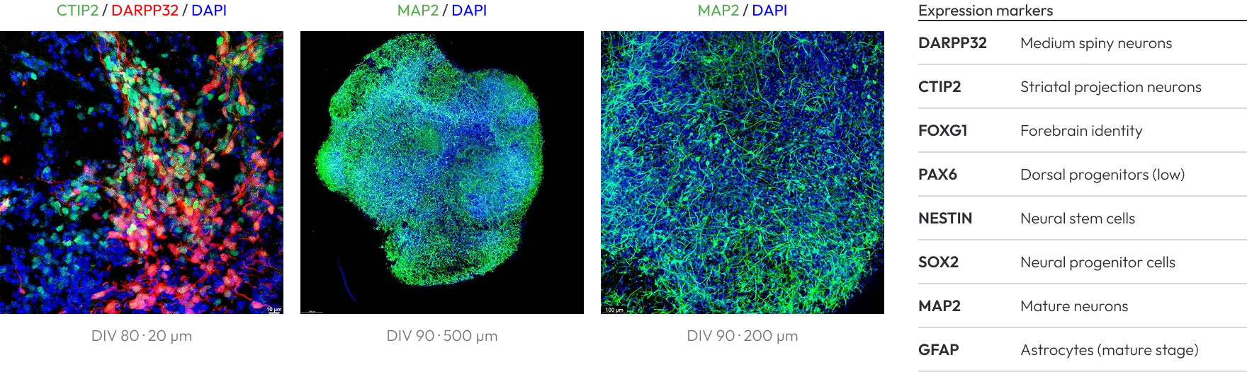
Striatal organoids recapitulate key features of the developing human striatum, including the generation and maturation of medium spiny neurons (MSNs), as demonstrated by the co-expression of DARPP32 and CTIP2. The organoids show low PAX6 and high FOXG1 expression, consistent with forebrain identity and striatal differentiation. GFAP expression becomes detectable in mature striatal organoids, indicating the emergence of astroglial populations during maturation. Together, these characteristics provide a physiologically relevant in vitro model for studying human striatal development, neuronal maturation, and striatum-associated neurological disorders.
Neural progenitors and forebrain identity (DIV 20)
Medium spiny neuron marker expression (DIV 45)
Mature striatal organoids (DIV 80–90)

Calcium Imaging of Functional Striatal Neurons

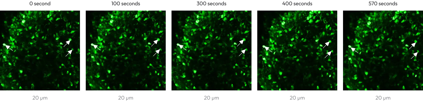
Beyond marker expression, striatal organoids display spontaneous neuronal activity. Live calcium imaging with the Fluo-4 AM indicator captures dynamic intracellular calcium signaling in individual cells over a 600-second recording, providing a functional readout for neuronal activity, drug response, and neurotoxicity assessment.
Fluo-4 AM time-lapse imaging (DIV 90)

Figure 1. Representative time-lapse fluorescence images of human striatal organoids loaded with the Fluo-4 AM calcium indicator. Selected cells exhibit variations in fluorescence intensity during the 600-second imaging period, indicating dynamic intracellular calcium signaling.

Frequently Asked Questions about Striatal Organoids

Striatal organoids are three-dimensional brain organoids derived from human induced pluripotent stem cells (iPSCs) that model the striatum, the forebrain region that controls movement, motivation, and reward. They contain medium spiny neurons (MSNs) and, as they mature, astroglial cells, providing a human-relevant model of striatal development and disease.
Human pluripotent stem cells are first aggregated into embryoid bodies (DIV -1), then guided through neural induction (DIV 0), striatal patterning and neural expansion (DIV 6), striatal differentiation (DIV 20), and striatal maturation (DIV 45 onward). Organoids can be maintained beyond DIV 100 for long-term maturation studies.
Medium spiny neurons (MSNs) are GABAergic projection neurons that make up the large majority of neurons in the human striatum. They are the cell type most affected in Huntington’s disease and a core part of the motor circuits disrupted in Parkinson’s disease, making them a key target for neurological drug discovery.
Immunostaining confirms co-expression of the MSN markers DARPP32 and CTIP2, high FOXG1 with low PAX6 expression (forebrain and striatal identity), NESTIN and SOX2 in early neural progenitors, MAP2 in mature neurons, and GFAP in astroglia of mature organoids. Neuronal function is assessed by Fluo-4 AM calcium imaging.
Striatal organoids are used for neurological drug efficacy testing, organoid-based neurotoxicity screening, assessment of neuronal function, and disease modeling of Huntington’s disease, Parkinson’s disease, and other movement and neurodevelopmental disorders.
Yes. Striatal organoids can be genetically modified, for example by introducing disease-associated mutations, to investigate the mechanisms underlying Huntington’s disease, Parkinson’s disease, and other movement disorders directly in human neurons.
Animal models do not fully recapitulate human brain biology, and 2D neuron cultures lack tissue architecture. Striatal organoids are human 3D tissues with self-organized structure and multiple cell types, providing more predictive data on human drug response and neurotoxicity.
Striatal differentiation begins around DIV 20 and maturation from DIV 45, with mature MSN populations characterized at DIV 80–90. Organoids can be cultured beyond DIV 100. Exact project timelines depend on your study design and are confirmed in your quote.
Pricing depends on your study design, including the number of organoids, culture duration, and the assays required. Request a quote to receive a tailored proposal for your project. Prefer to run the work in your own lab? We also offer striatal organoid culture media together with an optimized, validated protocol, so you can generate and maintain striatal organoids in-house with consistent results.

Thank you for your submission.

Our team has received your request and will get back to you shortly with tailored workflows and relevant case studies for your ADC efficacy and toxicity evaluation needs.
If you do not receive our confirmation email, kindly check your spam or junk folder.

Thank you for your interest

You can now download the file.

Connect with Us