Patient-derived organoids (PDOs) are increasingly used in drug discovery to evaluate efficacy, investigate mechanisms of action, and compare therapeutic response across heterogeneous patient-derived models.
But what does it actually mean for an organoid to be validated?
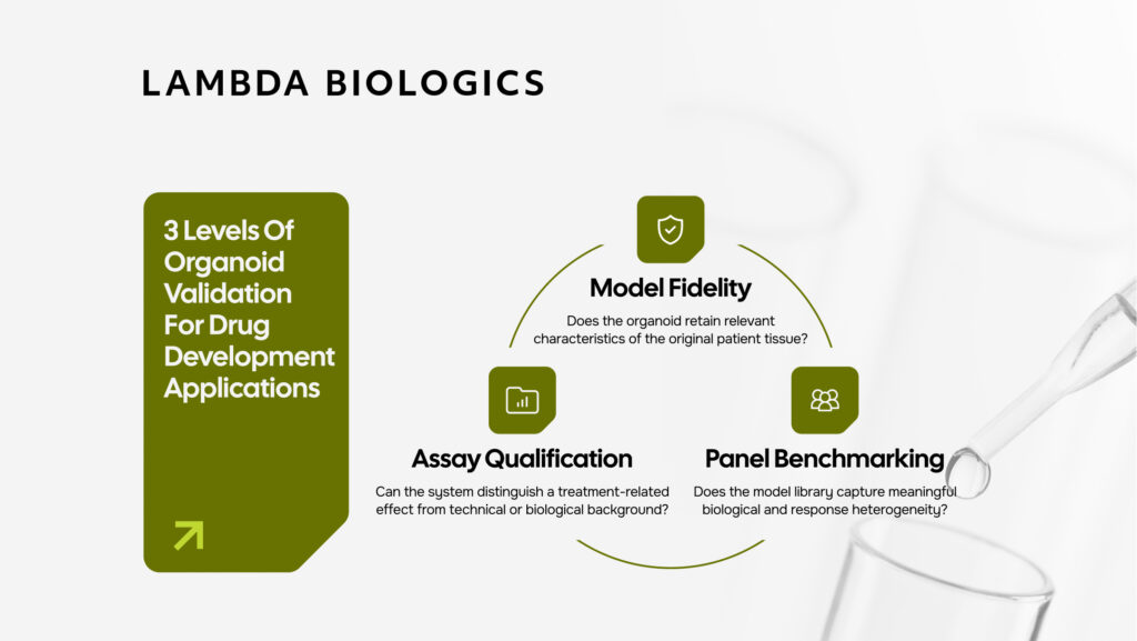
Morphology, immunohistochemistry, and genomic characterization are important, but they answer only one part of the question. A biologically characterized organoid does not automatically guarantee that the drug-response assay built around it is reliable. For drug-development applications, organoid validation should be considered at three levels:
- Model fidelity: Does the organoid retain relevant characteristics of the original patient tissue?
- Assay performance: Can the system distinguish a treatment-related effect from technical or biological background?
- Panel diversity: Does the model library capture meaningful biological and response heterogeneity?
This blog will explore how these three levels provide a practical framework for evaluating patient-derived organoid models and their suitability for drug testing at Lambda Biologics. To illustrate how these principles translate into practice, this article also includes a colorectal cancer (CRC) organoid case study using Lambda Biologics’ own characterization and validation data. The case study shows how multiple layers of evidence – from histology and molecular profiling to functional drug-response testing – can be combined to assess whether a PDO model is fit for its intended experimental use.
Read more: From Cell Lines to Patients: How CRC Organoids Strengthen Drug Development Workflows
In This Article
Model Fidelity: Does the Organoid Retain the Original Tumor Biology?
The first step is to establish whether the derived organoid retains key characteristics of the patient’s tumor. Rather than relying on a single measurement, characterization should combine complementary biological readouts.
Morphology and phenotype
H&E staining enables direct comparison between the parent tumor and derived organoid, while immunohistochemistry confirms tissue lineage and disease-relevant marker expression. Marker panels are selected according to tumor type and study purpose. Importantly, characterization should be performed at a defined passage, because phenotype and marker expression may change during prolonged culture.
Genomic concordance
Where possible, the original tumor and derived organoid should also be analyzed as a matched pair. This allows researchers to determine whether relevant driver alterations are retained instead of assuming genomic fidelity based on published organoid studies.
Differences in variant allele frequency may occur because organoid establishment enriches the epithelial tumor population and removes much of the stromal and immune-cell background present in bulk tissue. The key question is therefore not whether every quantitative measurement remains identical, but whether biologically relevant genomic features remain detectable and interpretable.
Molecular phenotype
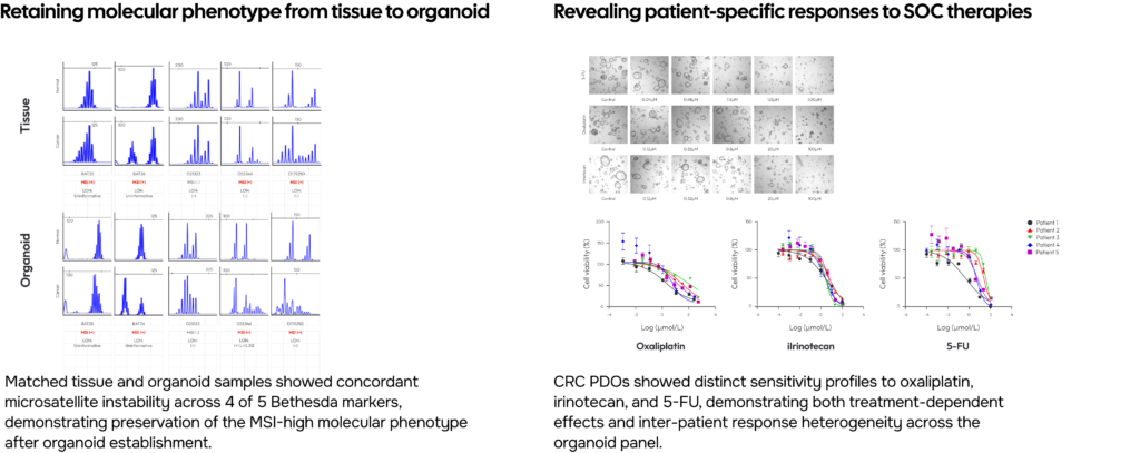
Disease-relevant molecular features can provide an additional validation layer. For colorectal cancer, for example, microsatellite status can be evaluated between tumor-derived material and matched normal material where available, helping determine whether the molecular phenotype is retained after organoid establishment.
Together, morphology, phenotype, genomic characteristics, molecular subtype, and passage information establish the biological fidelity of a PDO model. But this still does not establish whether the assay itself is reliable.
Assay Qualification: Can the Experiment Distinguish a True Treatment Effect?
Once an organoid is used for efficacy, toxicity, or immune co-culture studies, additional variables can affect the result. Assay qualification therefore requires appropriate controls and measurable acceptance criteria.
These variables include:
- organoid number and size;
- plating variability;
- direct compound toxicity;
- nonspecific immune-cell killing;
- effector-to-target ratio;
- assay duration;
- passage and culture conditions.
Baseline uniformity
Organoid count and size should be assessed before treatment to ensure that experimental groups begin from comparable conditions. Without this control, apparent differences after treatment may partly reflect variation introduced during plating.
Drug-only controls
In immune-mediated studies, the drug should also be tested against the organoid without immune effector cells. This helps distinguish direct drug toxicity from immune-cell-dependent activity.
Background and nonspecific killing
Immune co-culture inherently introduces biological background. Depending on the study, this can be evaluated using controls such as:
- untreated co-culture;
- isotype controls;
- organoid-only conditions;
- donor-mismatched immune cells;
- viability measurements.
These controls establish the background against which treatment-dependent activity is interpreted.
They can also help define an assay-specific acceptance threshold, below which an observed effect should not be considered meaningful.
Specificity and assay window
For immune-mediated therapies, matched and non-matched target systems can provide strong evidence that killing is dependent on the intended biological interaction rather than generic cytotoxicity.
Assay performance should also be evaluated across relevant conditions, such as effector-to-target ratios and timepoints.
Variables such as immune-cell age or organoid size may alter assay sensitivity. These conditions therefore need to be defined as part of the experimental protocol rather than treated as operational details.
The objective is to establish a measurable window in which the assay can reliably detect the intended biological effect.
Read more: Cancer Organoid Co-culture with Macrophages and T Cells for Drug Evaluation
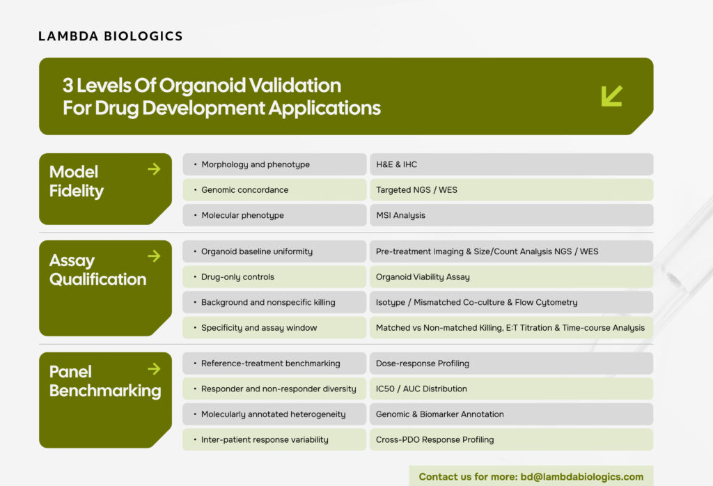
Panel Benchmarking: Does the Model Library Capture Biological Heterogeneity?
A reproducible assay is only as informative as the biological diversity of the models tested. For screening studies, an organoid panel should contain enough variation to capture sensitive, intermediate, and resistant phenotypes.
One way to assess this is by benchmarking individual models against reference compounds or standard-of-care treatments. A meaningful PDO panel should demonstrate a distribution of drug responses, rather than uniformly high or uniformly low sensitivity.
Molecular annotation adds another layer of value. Models can be stratified according to characteristics such as:
- tumor subtype;
- anatomical origin;
- microsatellite status;
- driver mutations;
- biomarker expression;
- therapeutic-target expression.
This allows researchers to ask not only whether a candidate works, but also in which biological context it works best. Inter-patient response variability is therefore not simply experimental noise. When supported by appropriate assay controls, it is one of the principal advantages of using patient-derived organoid panels.
Case Study: Validation of Colorectal Cancer Organoids
Colorectal cancer provides a useful example of how these validation principles can be applied in practice. At Lambda Biologics, colorectal cancer organoids can be characterized through multiple complementary layers before being used in drug-evaluation studies.
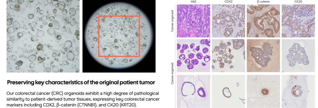
Parent tumor tissue and corresponding PDOs can be compared using H&E and colorectal lineage markers.
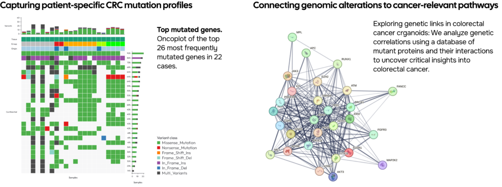
Colorectal cancer is a highly heterogeneous disease, requiring models that accurately reflect the diversity of patient tumors. Our CRC organoid bank comprises genomically characterized, patient-derived organoids that enable mutation-specific model selection for drug testing, biomarker discovery, mechanism-of-action studies, and resistance research.
Drug sensitivity tests have been conducted using 5-FU, Oxaliplatin, and Irinotecan which are widely used in clinical cancer treatment, and drug response data has been collected from a subset of CRC organoid models, comprising over 20 distinct lines.
Toward Fit-for-Purpose Organoid Validation
There is no single measurement that can determine whether an organoid is suitable for every drug-development application. Different studies require different evidence: A model used for small-molecule screening may require different qualification criteria from an organoid–immune-cell co-culture used to evaluate an immunotherapy.
For this reason, organoid validation should move beyond a generic characterization checklist toward fit-for-purpose qualification (FDA Modernization Act 3.0) that incorporates biological fidelity, assay-specific controls, and functional benchmarking. At Lambda Biologics, the objective is not simply to demonstrate that an organoid has been successfully established. It is to answer a more useful question: Can this model, under these experimental conditions, generate reliable data for the biological question being asked? That is ultimately what organoid validation should demonstrate.
Frequently Asked Questions
How are patient-derived organoids validated?
Which assays are commonly used to validate cancer organoids?
How is tumor–organoid genomic concordance assessed?
What controls are required to qualify an organoid–immune cell killing assay?
How are assay acceptance criteria defined for organoid drug-response studies?
Planning a Human-Relevant Efficacy or Toxicity Study?
Talk to Lambda Biologics about an organoid-based study designed around your molecule, biological question and development objectives.
Contact us to discuss your research!
Lambda Biologics’s Organoid-based Discovery platform for Innovative Screening, Evaluation, and Identification