Despite aggressive treatments like chemotherapy, cytoreductive surgery (CRS), and hyperthermic intraperitoneal chemotherapy (HIPEC), peritoneal metastases (PM) remain a common and challenging site of disease progression in colorectal cancer (CRC), often leading to poor outcomes.
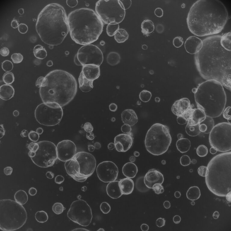
Researchers have utilized Matrix-Assisted Laser Desorption/Ionization – Time of Flight (MALDI-TOF) mass spectrometry (MS) and imaging (MALDI-IMS) to investigate the lipid profiles in patient-derived tumor organoids (PDOs) and PM samples. Their study revealed that the predominant lipid signature in PM tissues is glycosphingolipid (GSL) sulfates or sulfatides (STs), which are unique to tumor areas and absent in surrounding stromal tissue.
The stromal components predominantly express bioactive lipids derived from arachidonic acid (AA) and specific phosphatidylinositol (PI) species, whereas tumor components contain PI species with shorter, more saturated acyl chains. These lipid alterations may facilitate tumor growth, aggressiveness, and metastasis.
The study suggests that targeting the GSL/ST metabolic pathways in PM could present new therapeutic opportunities to slow or stop disease progression.
Keywords: sulfatide imaging, cancer, metastasis, colorectal cancer


