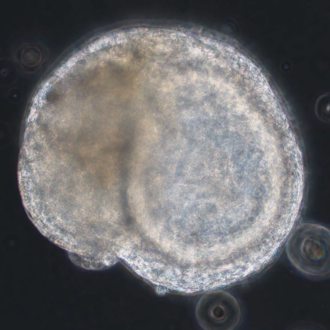
Our cardiac organoids recapitulate fundamental aspects of human cardiac function, exhibiting spontaneous and rhythmic contractility that closely mirrors native myocardial activity. These physiologically relevant 3D models provide a robust platform for investigating cardiac development, disease mechanisms, and drug-induced cardiotoxicity.
Price | 5500€+ |
Organism | Human |
Product Type | iPSC-derived organoid |
Tissue | Cardiac |
Disease | – |
Applications
Organoid Based Toxicity

With the global rise of K-beauty, the cosmetics industry continues to grow steadily. Since the ban on animal testing for cosmetics in Korea in 2017, various alternative testing methods have...

Traditional microscopy methods often require fluorescent labeling to analyze cellular structures, which can be time-consuming and invasive. In contrast, our HT-X1 system allows for high-resolution visualization of cellular morphology without...

Traditional protein analysis has primarily focused on quantifying expression levels within tissue samples. However, recent advances in spatial analysis techniques have shifted attention toward evaluating not only expression levels, but...
Among the many fermented foods we consume, kimchi is particularly known for containing a diverse range of lactic acid bacteria, which are believed to influence the activation of immune cells...
We conducted a study focused on identifying disease-related markers using patient-derived tissue samples. However, traditional methods limited our ability to analyze multiple candidate markers simultaneously, and the limited availability of...
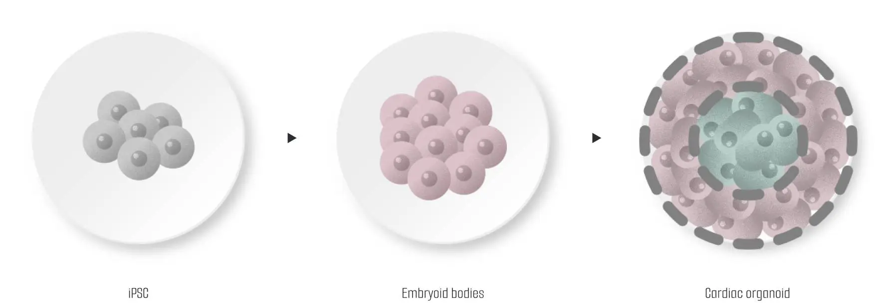
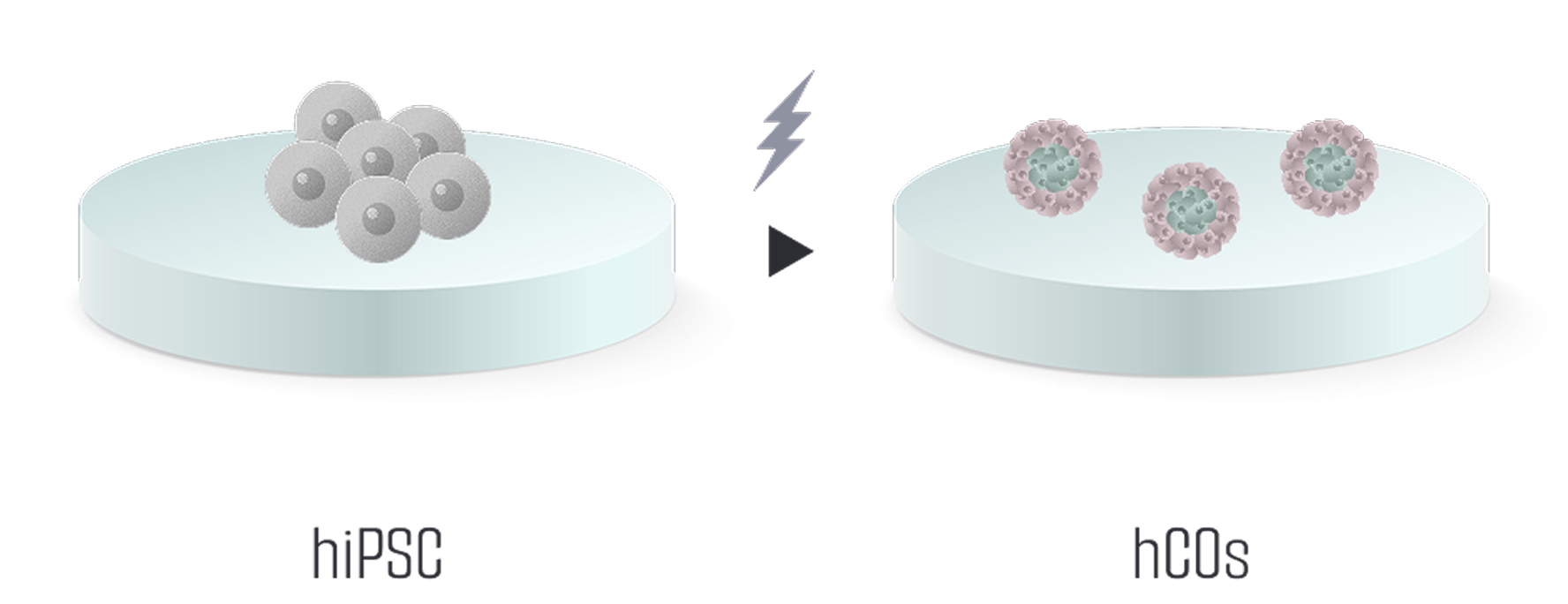
Our cardiac organoids are engineered from human-induced pluripotent stem cells (iPSCs). By transforming 3D embryoid bodies through precise signal manipulation, we create organoids that emulate heart tissue, advancing personalized cardiac research and therapy.
The cultivation process has been simplified by eliminating extracellular matrices, reducing variability and improving the efficiency of organoid production. This streamlined approach ensures more consistent and reproducible results in a shorter timeframe.
Through refined differentiation techniques, we achieve superior maturation of the cardiac organoids, carefully balancing myocardial and non-myocardial cells to replicate the complex structure of the human heart. This enhanced maturation leads to more accurate and functional models for research applications.


– Cardiomyocytes
– Non-myocytes
(cardiac fibroblast, endothelial cells)

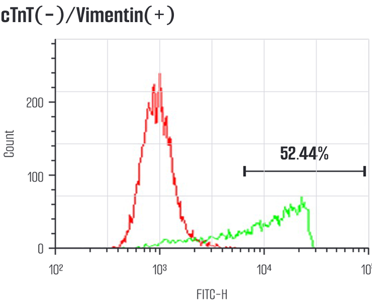
The cardiac organoids produced are composed similarly to the human heart, containing cardiomyocytes and non-muscle cells like cardiac fibroblasts and endothelial cells.
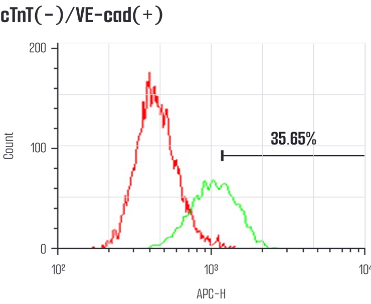
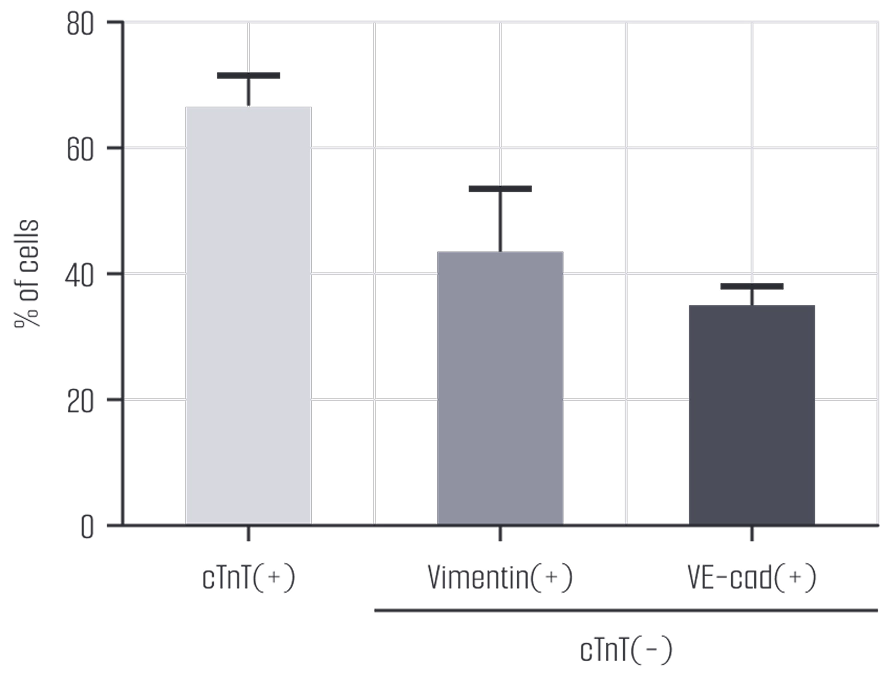
Through FACS analysis, it was determined that cardiomyocytes constitute about 65% of the cell population, with the majority of the non-muscle cells being fibroblasts and endothelial cells.


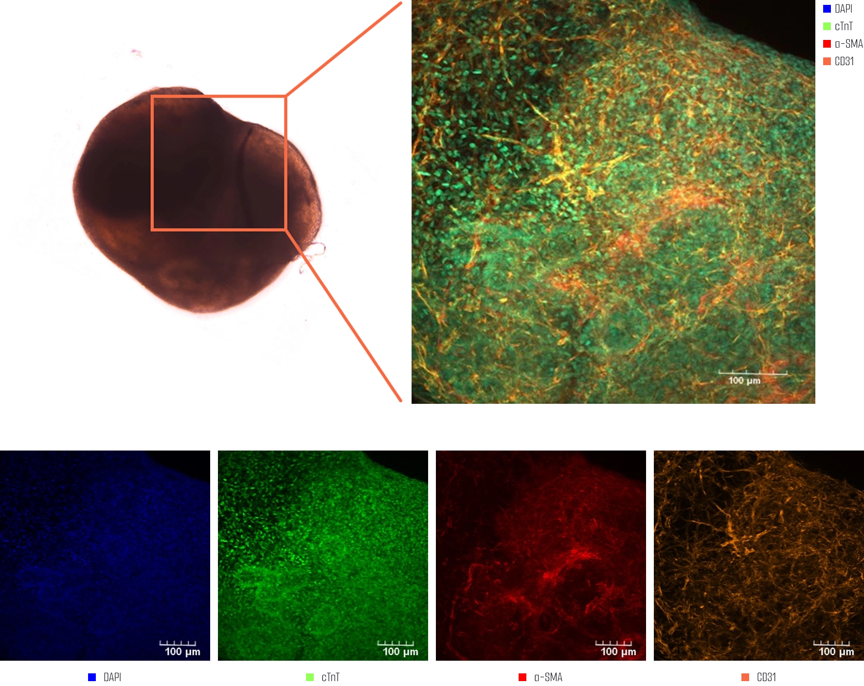
Using immunofluorescence, we have confirmed the expression of myocardial markers (cTnT, α-actinin), fibroblast markers (α-SMA), and endothelial cell markers (CD31) in our cardiac organoids.
This method allows for detailed visualization and verification of specific cellular components critical to heart tissue function.


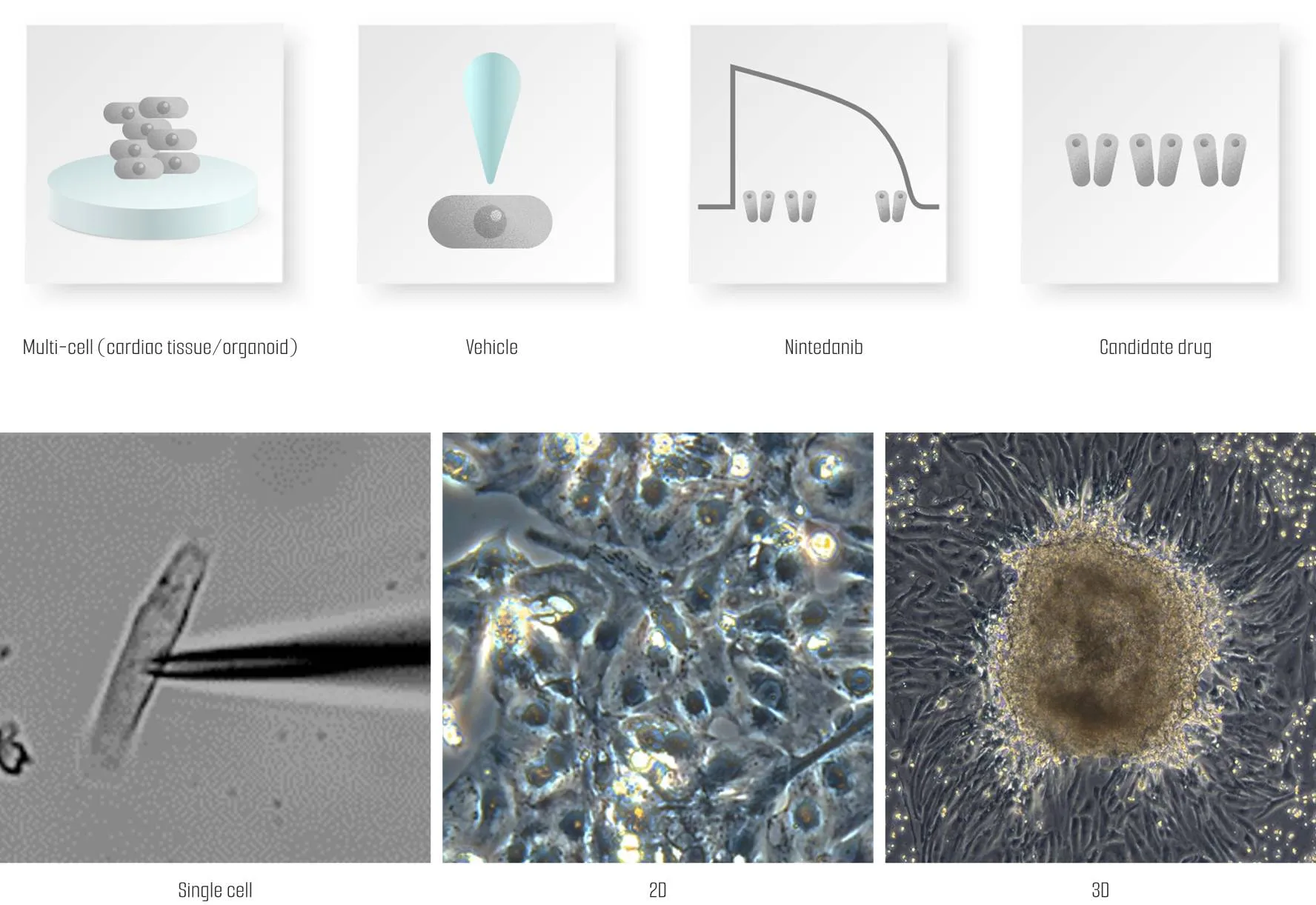
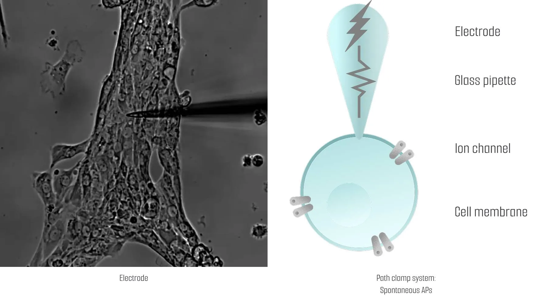
To analyze the electrophysiological properties of cardiac organoids with spontaneous beating, we employ the patch clamp technique. By inserting electrodes into the cell membranes of the cardiac organoids, we can monitor action potentials in real-time, providing valuable insights into their functional behavior.
Explore how cardiac organoids can support your electrophysiological studies.
Contact Lambda Biologics to discuss your project with our team.
Cardiac organoids are three-dimensional, self-organizing tissues derived from human pluripotent stem cells. Unlike 2D monolayer cultures, they contain multiple cardiac cell types and exhibit spontaneous rhythmic contractions, enabling more physiologically relevant modeling of heart development and function.
They are applied in cardiotoxicity screening, disease modeling (e.g., cardiomyopathies, myocarditis), cardiac development studies, and regenerative medicine research. Their human-relevant physiology also supports early-stage drug efficacy and safety assessment.
Our organoids are generated using a matrix-free, standardized cultivation process that minimizes variability and supports consistent batch production. This streamlined approach ensures reproducible functional and structural outcomes across experiments.
Through refined differentiation and culture optimization, the organoids display advanced maturation, featuring coordinated contractility, electrophysiological activity, and a balanced representation of myocardial and non-myocardial cells.
Yes. We offer customization based on donor background, disease-specific iPSC lines, or assay compatibility to align with individual project requirements, from mechanism-of-action studies to compound screening.