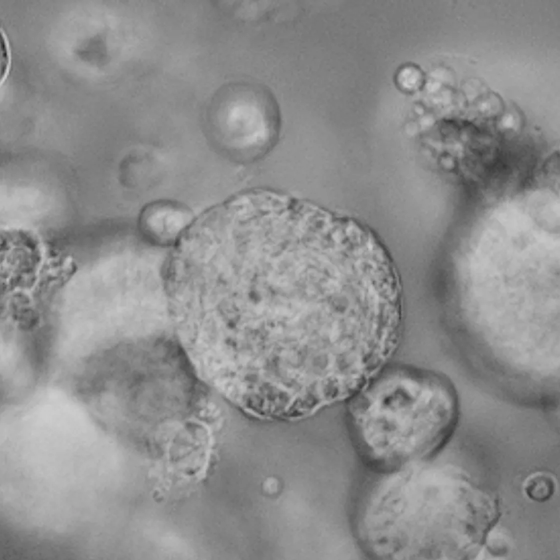
Lambda Biologics’ lacrimal gland organoids faithfully replicate native tear-gland tissue - matching histology and key marker expression, and demonstrating pilocarpine-induced Ca²⁺ signaling with β-hexosaminidase release. Enable more predictive drug screening, disease modeling, and regenerative studies with a system that captures true lacrimal function.
Price | 4850€+ |
Organism | Human |
Product Type | Adult Tissue derived organoid |
Tissue | Lacrimal gland |
Disease | Gland Disease Model |
Applications
Organoid Based Toxicity

With the global rise of K-beauty, the cosmetics industry continues to grow steadily. Since the ban on animal testing for cosmetics in Korea in 2017, various alternative testing methods have...

Traditional microscopy methods often require fluorescent labeling to analyze cellular structures, which can be time-consuming and invasive. In contrast, our HT-X1 system allows for high-resolution visualization of cellular morphology without...

Traditional protein analysis has primarily focused on quantifying expression levels within tissue samples. However, recent advances in spatial analysis techniques have shifted attention toward evaluating not only expression levels, but...
Among the many fermented foods we consume, kimchi is particularly known for containing a diverse range of lactic acid bacteria, which are believed to influence the activation of immune cells...
We conducted a study focused on identifying disease-related markers using patient-derived tissue samples. However, traditional methods limited our ability to analyze multiple candidate markers simultaneously, and the limited availability of...
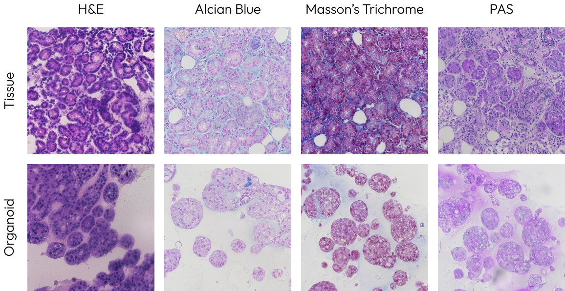
In the lacrimal gland organoid, the structure of acinar cells and their secretion of acidic mucins were observed by H&E, Alcian blue. Additionally, through PAS staining, secretions such as glycogen and glycoproteins were observed in the organoid. Masson’s Trichrome staining demonstrated that keratin is a major ECM component in the organoid.

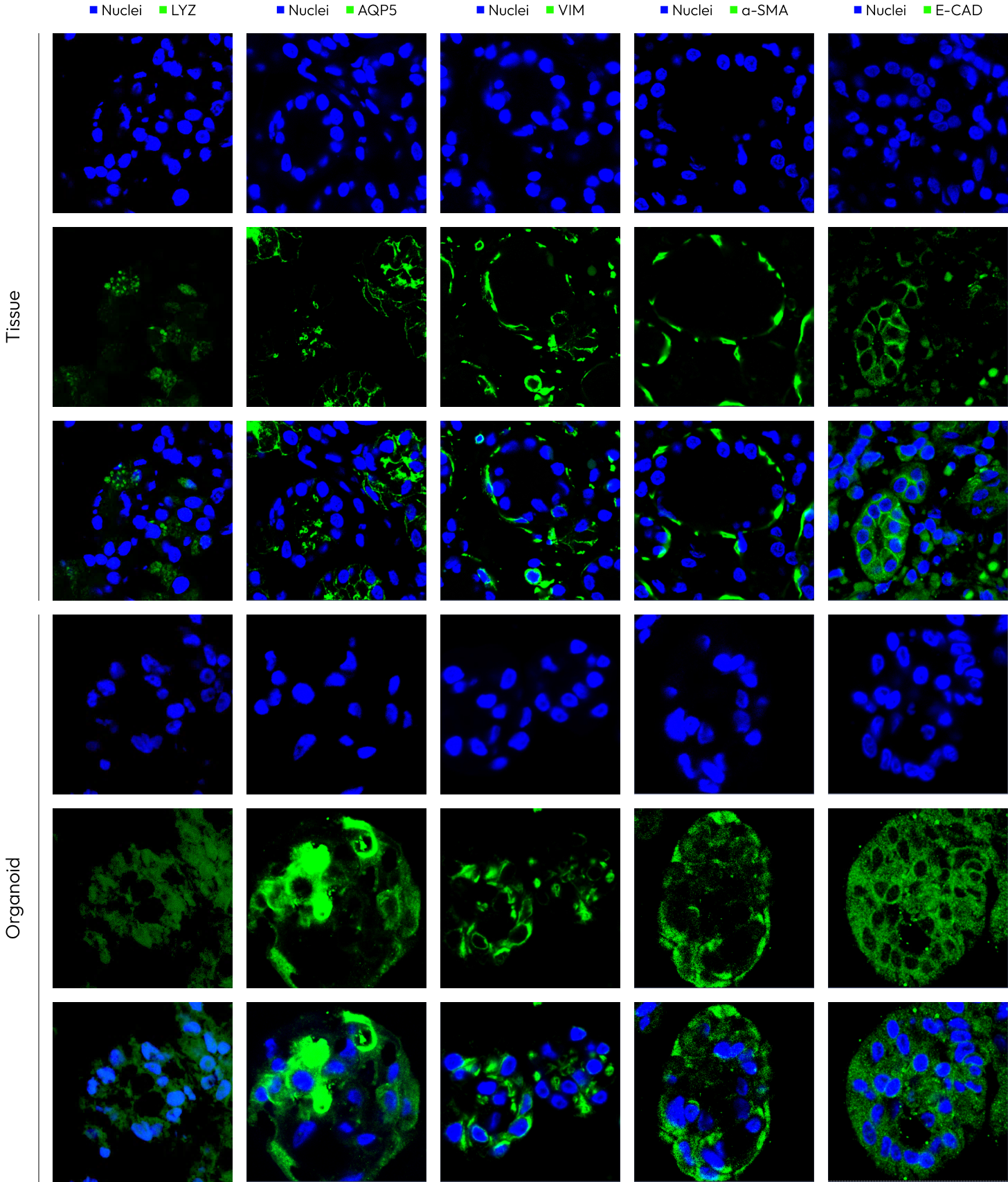
The expression patterns of VIM, AQP5, and LYZ within the organoid closely resemble those of the tissue’s acinar cells. Furthermore, the expression patterns of E-CAD and α-SMA in the organoid are similar to those in the tissue, though at lower levels. These markers are not expressed in ductal cells but are expressed in acinar cells, confirming that the formed organoid closely resembles acinar cells.

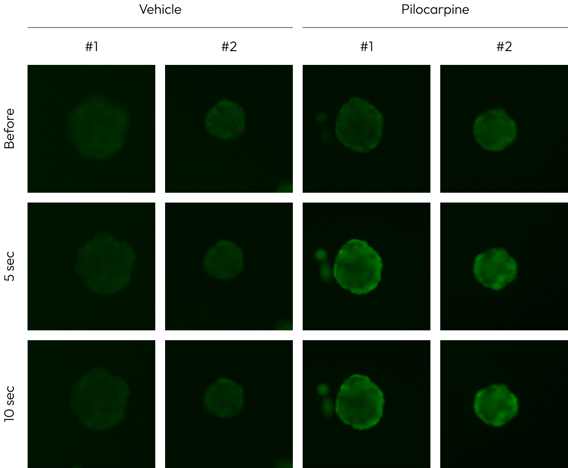
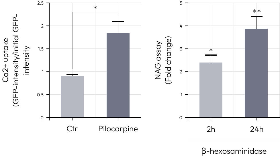
To assess the secretory function, lacrimal gland organoids were treated with pilocarpine.
The intracellular concentration of Ca ions increased, confirming the elevation of the lysosomal protein β-hexosaminidase, a component of tears.


Interested in bringing lacrimal gland organoids into your research?
Contact Lambda Biologics to discuss your project with our team.
Our 3D organoids recapitulate native lacrimal gland architecture, express key tear-gland markers, and respond functionally to pilocarpine. This provides a more predictive platform than 2D cultures for studying secretion, disease mechanisms, and drug effects.
They are ideal for dry-eye disease modeling, drug efficacy and toxicity testing, tear-secretion studies, and exploring regenerative or cell-therapy approaches for lacrimal gland repair.
We provide detailed technical data sheets covering differentiation methods, marker expression, and functional validation, so you can trust the reliability and reproducibility of your results.
Yes. Lambda Biologics offers flexible formats – frozen organoids, live cultures, or assay-ready plates – and can collaborate on protocol adaptation to fit your specific experimental design.
Simply contact Lambda Biologics team. Our scientists will review your project needs and provide the right options whether it’s technical guidance, a quote, or custom organoid development.