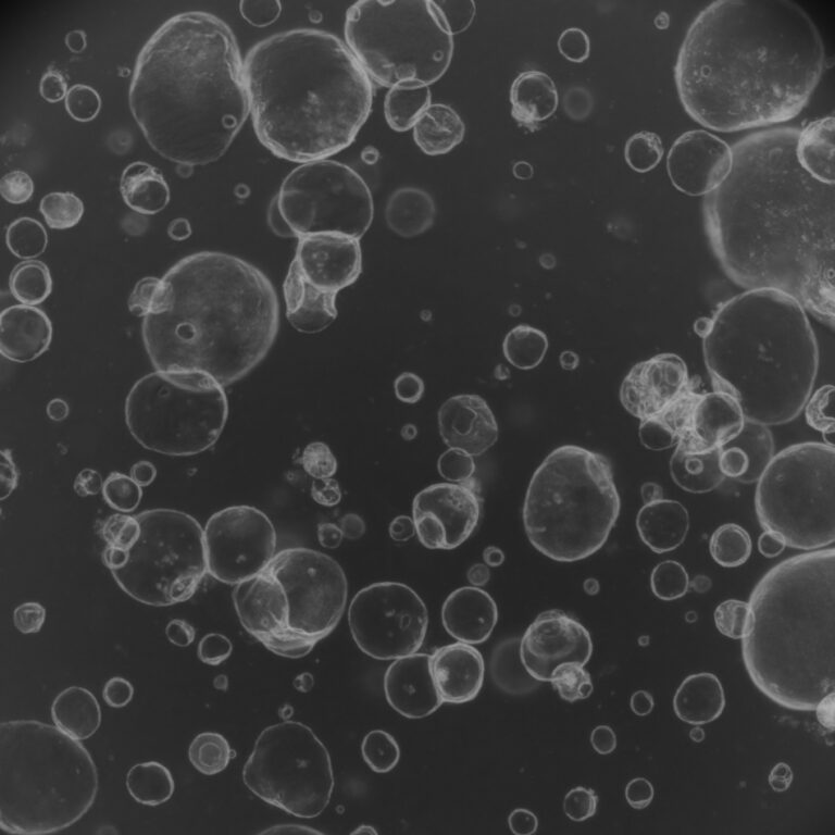
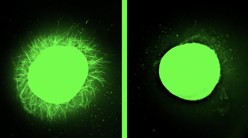
Johns Hopkins researchers have developed a pioneering multi-region human brain organoid – a lab-grown mini‑brain that incorporates neural tissues from the forebrain, midbrain, and hindbrain, along with rudimentary blood vessels. By separately culturing neural cells from each brain region and nascent vascular cells, then combining them using sticky proteins acting as “biological superglue,” the tissues fused into a unified organoid. As the composite grew, it began producing electrical signals and functioning as an integrated network – closely simulating early human brain dynamics.
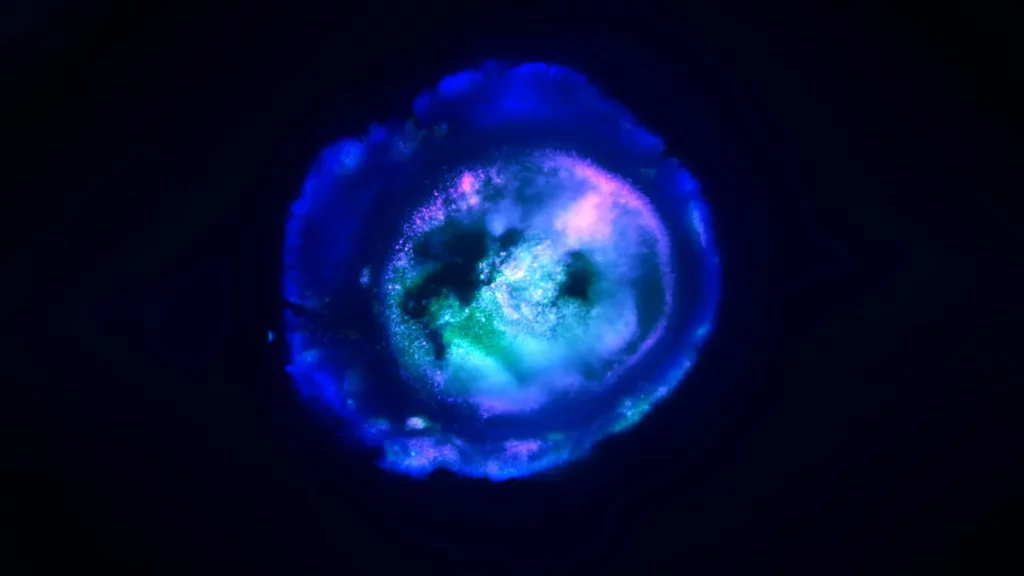
The organoid contains approximately 6 – 7 million neurons, significantly fewer than the tens of billions in an adult human brain, but sufficient to replicate characteristics of a ~40-day-old human fetus. Importantly, MRBOs exhibit about 80% of the cell types found in early fetal brain development stages. Researchers also detected emergent blood–brain barrier features, such as endothelial layers that regulate molecular entry into brain-like tissue.
Lead author Annie Kathuria emphasized that MRBOs allow scientists to observe neurodevelopmental disorders in real time and evaluate treatments using human cells – bypassing limitations of animal-based models. The organoid platform offers a human-relevant system to explore conditions like schizophrenia, autism, and Alzheimer’s disease, and to test drugs and therapies – potentially lowering the high failure rate (≈96%) seen in neuropsychiatric drug trials.
Research article: Johns Hopkins scientists grow a mini human brain that lights up and connects like the real thing
Lambda Biologics’ Oncology Solutions: Patient-derived cancer organoid-based drug evaluation service
Gastric Cancer Organoid | Breast Cancer Organoid | Hepatocarcinoma Cancer Organoid | Pancreatic Cancer Organoid
