
Salivary gland organoid closely resemble native salivary gland tissue - both in structure and function. Featuring key cell types, including acinar and ductal cells, these organoids provide a reliable platform for studying secretion, disease mechanisms, and regenerative therapies.
Price | 2850€+ |
Organism | Human |
Product Type | Adult Tissue derived organoid |
Tissue | Salivary Gland |
Disease | Gland Disease Model |
Applications
Organoid Based Toxicity

With the global rise of K-beauty, the cosmetics industry continues to grow steadily. Since the ban on animal testing for cosmetics in Korea in 2017, various alternative testing methods have...

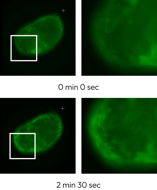
Traditional microscopy methods often require fluorescent labeling to analyze cellular structures, which can be time-consuming and invasive. In contrast, our HT-X1 system allows for high-resolution visualization of cellular morphology without...

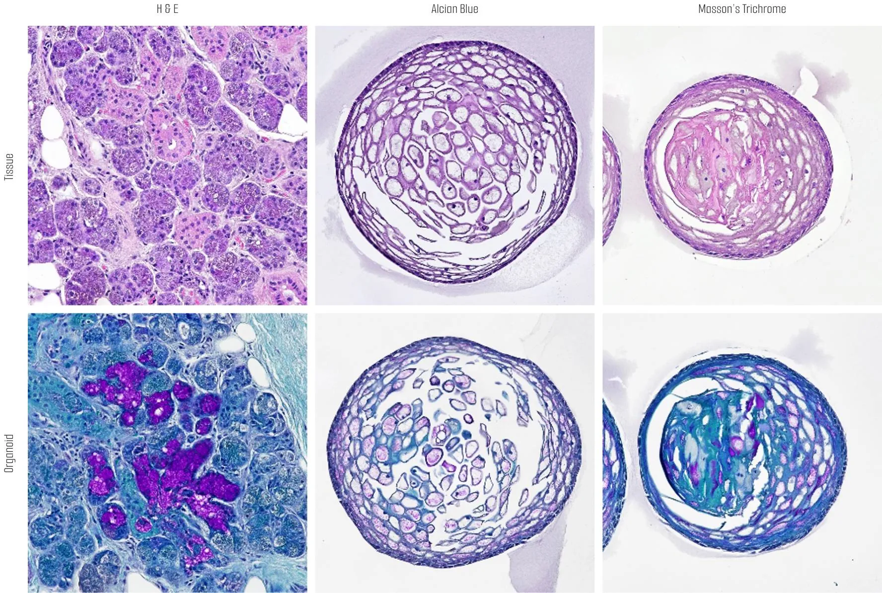
Traditional protein analysis has primarily focused on quantifying expression levels within tissue samples. However, recent advances in spatial analysis techniques have shifted attention toward evaluating not only expression levels, but...
Among the many fermented foods we consume, kimchi is particularly known for containing a diverse range of lactic acid bacteria, which are believed to influence the activation of immune cells...
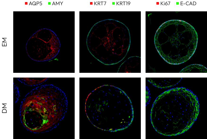
We conducted a study focused on identifying disease-related markers using patient-derived tissue samples. However, traditional methods limited our ability to analyze multiple candidate markers simultaneously, and the limited availability of...
Histological analyses (H&E, PAS) demonstrate enhanced cellular complexity and a rise in mucin-producing cells as the organoids undergo differentiation. These findings confirm that tissue-level similarity strengthens during the differentiation process, reinforcing their value as human-relevant models for drug discovery and regenerative research.


Immunofluorescence analysis reveals that differentiated organoids show elevated expression of acinar cell markers (AQP5, AMY) and a polarized distribution of ductal markers (CK7, CK19), closely mirroring the structural and functional organization of native salivary gland ducts.
The ability to secrete saliva was verified by observing the response of calcium accumulation upon stimulation with the cholinergic agonist carbachol.

Explore how salivary gland organoids can support your research while reducing costs and ethical barriers. Connect with our experts to learn more.
Our organoids faithfully mimic the 3D architecture and cellular composition of human salivary glands. Histological and immunofluorescence analyses show key acinar and ductal markers (AQP5, AMY, CK7, CK19) and demonstrate polarized organization comparable to natural ducts, ensuring a physiologically relevant model for disease and functional studies.
Yes. We can adjust differentiation stage, culture conditions, and marker expression to meet diverse objectives – from high-throughput drug testing to regenerative medicine and disease modeling. This flexibility supports both basic research and translational projects.