Meet us at ACS FALL 18 - 20 August
ODISEI Organoid-based Discovery Platform Introduction
Meet us at ACS FALL 18 - 20 August
ODISEI Organoid-based Discovery Platform Introduction
Meet us at ACS FALL 18 - 20 August
ODISEI Organoid-based Discovery Platform Introduction
Meet us at ACS FALL 18 - 20 August
ODISEI Organoid-based Discovery Platform Introduction
Home » AFS » Midbrain Organoids
Midbrain Organoids
Organoid Service

Midbrain Organoids

9275€+

Midbrain Organoids

Lambda Biologics midbrain organoids give researchers a powerful, human-relevant model to study brain development and disease. By replicating the key structures and cell types of the human midbrain, they enable more accurate testing of drug efficacy and toxicity, reduce reliance on animal models, and deliver insights that translate more effectively to patient outcomes.

Price
9275€+
Organism
Human
Product Type
iPS-derived organoid
Tissue
Brain (Midbrain)
Disease
Parkinson’s Disease, Brain & CNS Disease Model

Applications

Toxicity

Organoid Based

Disease Modeling

Brain & CNS Disease Model

Professor Lee Chang-seok Eulji University
Customer insight

Advancing K-Beauty with Skin Organoids: A Next-Generation Platform for Non-Animal Testing and High-Precision Cosmetic Innovation

With the global rise of K-beauty, the cosmetics industry continues to grow steadily. Since the ban on animal testing for cosmetics in Korea in 2017, various alternative testing methods have...

Tomocube (Spatial)
Customer insight

HT-X1: A Label-Free Imaging Breakthrough for Organoid-Based Disease Modeling and Drug Screening

Traditional microscopy methods often require fluorescent labeling to analyze cellular structures, which can be time-consuming and invasive. In contrast, our HT-X1 system allows for high-resolution visualization of cellular morphology without...

Seoul National University College of Medicine
Customer insight

Pioneering Spatial Protein Analysis in Korea: Advancing Clinical Pathology with Lambda Biologics’ Support

Traditional protein analysis has primarily focused on quantifying expression levels within tissue samples. However, recent advances in spatial analysis techniques have shifted attention toward evaluating not only expression levels, but...

K Research Institute
Customer insight

ODISEI-Gut Platform Reveals Immune-Boosting Potential of Kimchi-Derived Bacterial Strain

Among the many fermented foods we consume, kimchi is particularly known for containing a diverse range of lactic acid bacteria, which are believed to influence the activation of immune cells...

Bundang Jesaeng General Hospital
Customer insight

Multiplex Marker Analysis Enhances Research Efficiency with 31-Marker Detection on a Single Slide

We conducted a study focused on identifying disease-related markers using patient-derived tissue samples. However, traditional methods limited our ability to analyze multiple candidate markers simultaneously, and the limited availability of...

Description

Generation Process

Through our proprietary differentiation technology, hPSC-derived midbrain organoids can be cultured long term and develop into advanced models enriched with mature dopamine neurons. These organoids offer a reliable system for investigating neurodevelopment and neurodegenerative diseases, including Parkinson’s.

Cellular and Structural Similarity

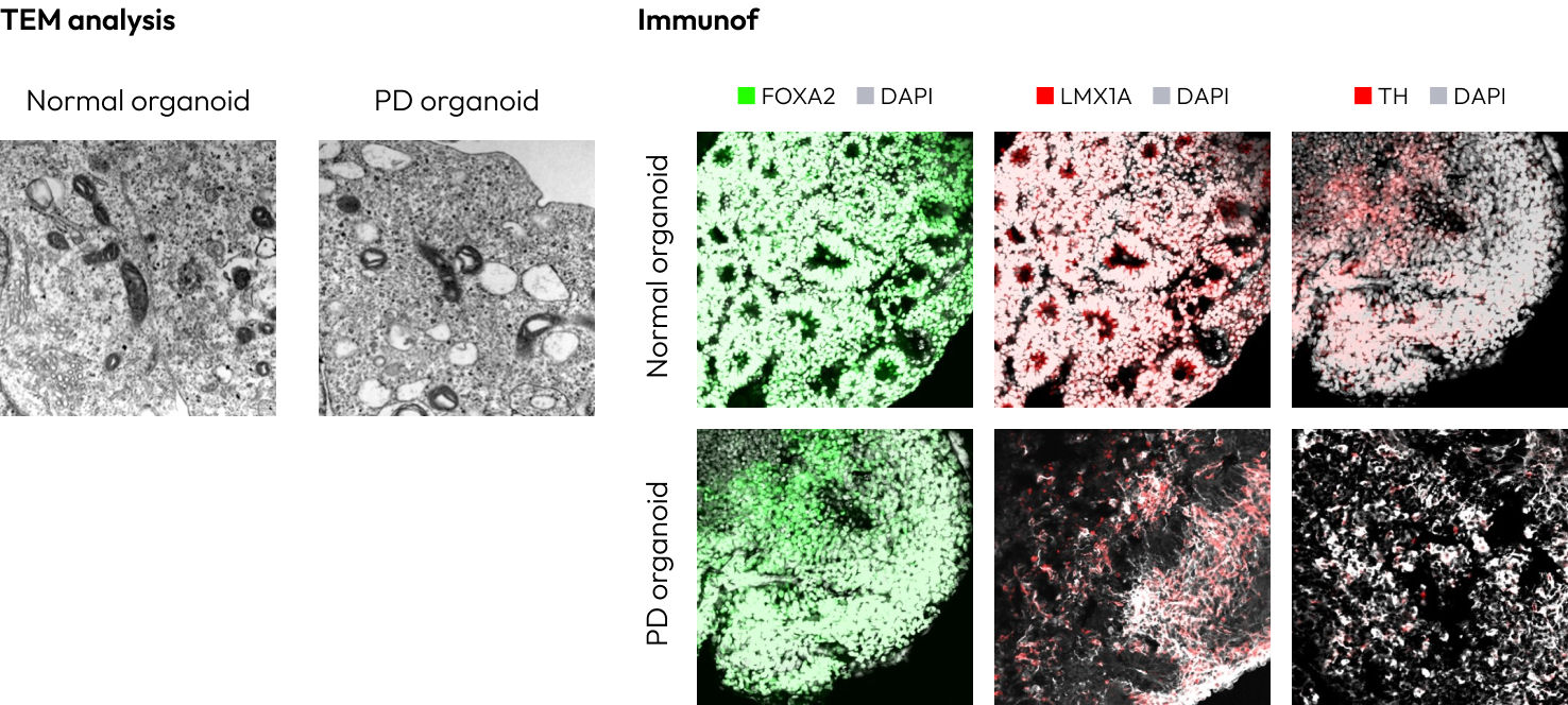
Our midbrain organoids replicate the key genetic and structural features of the human midbrain. They contain abundant dopamine neurons along with diverse supporting cell types. When cultured long term, they even develop melanin in the substantia nigra – a hallmark feature unique to the human midbrain.

FOXA2 – A transcription factor crucial for the development and differentiation of midbrain and hindbrain neurons, promoting the formation of midbrain dopaminergic neurons.
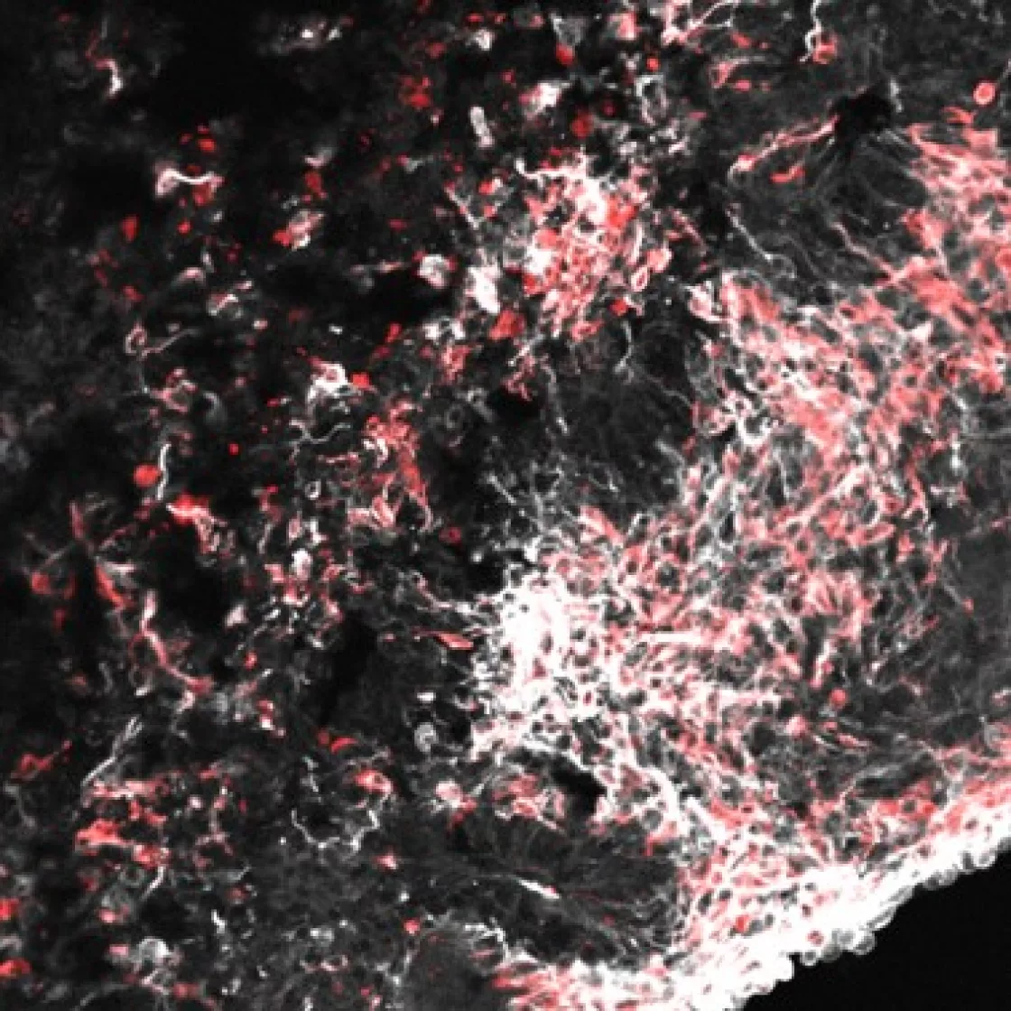
LMX1A – A transcription factor that regulates the differentiation of dopaminergic neurons in the midbrain and hindbrain.
TH (Tyrosine Hydroxylase) – An enzyme involved in dopamine synthesis, commonly used as a marker for dopaminergic neurons.
Nestin – A marker for neural progenitor cells and immature neurons, indicating immature cells in midbrain organoids.
OTX2 – A transcription factor important for the development of the forebrain and midbrain, expressed in the midbrain and brainstem regions.

Functional similarity

Our brain organoids exhibit electrical activity from nerve cells, reflecting patterns seen in the human brain. This confirms their functional maturity. With long term culture, they can even generate brain-wave patterns similar to those observed in premature infants.

Parkinson’s Disease Modeling

We can generate organoid models of neurodegenerative diseases through two methods: creating brain disease organoids using iPSCs with regulated expression of specific genes or inducing brain disease organoids through drug treatment.

Two main approaches are used to model Parkinson’s disease. The first involves utilizing gene editing technologies to modify or alter genes associated with Parkinson’s, such as introducing mutations in LRRK2 or SNCA to replicate disease-causing genes and analyze pathological features. The second approach involves creating organoids from patient-derived iPSCs (induced pluripotent stem cells).

By using iPSCs from Parkinson’s patients, personalized disease models can be developed, allowing researchers to experimentally recreate disease characteristics like dopaminergic neuronal degeneration and study drug development and therapeutic strategies.

Result

Parkinson’s disease is marked by the progressive loss of dopamine-producing neurons, driven in part by mitochondrial dysfunction. Mitochondrial swelling – a pathological enlargement of mitochondria – disrupts energy production and calcium balance, leading to cell death. This dysfunction also heightens oxidative stress and impairs intracellular signaling, ultimately damaging dopamine neurons. Together, these processes drive the motor symptoms of Parkinson’s disease, including tremors and movement difficulties.

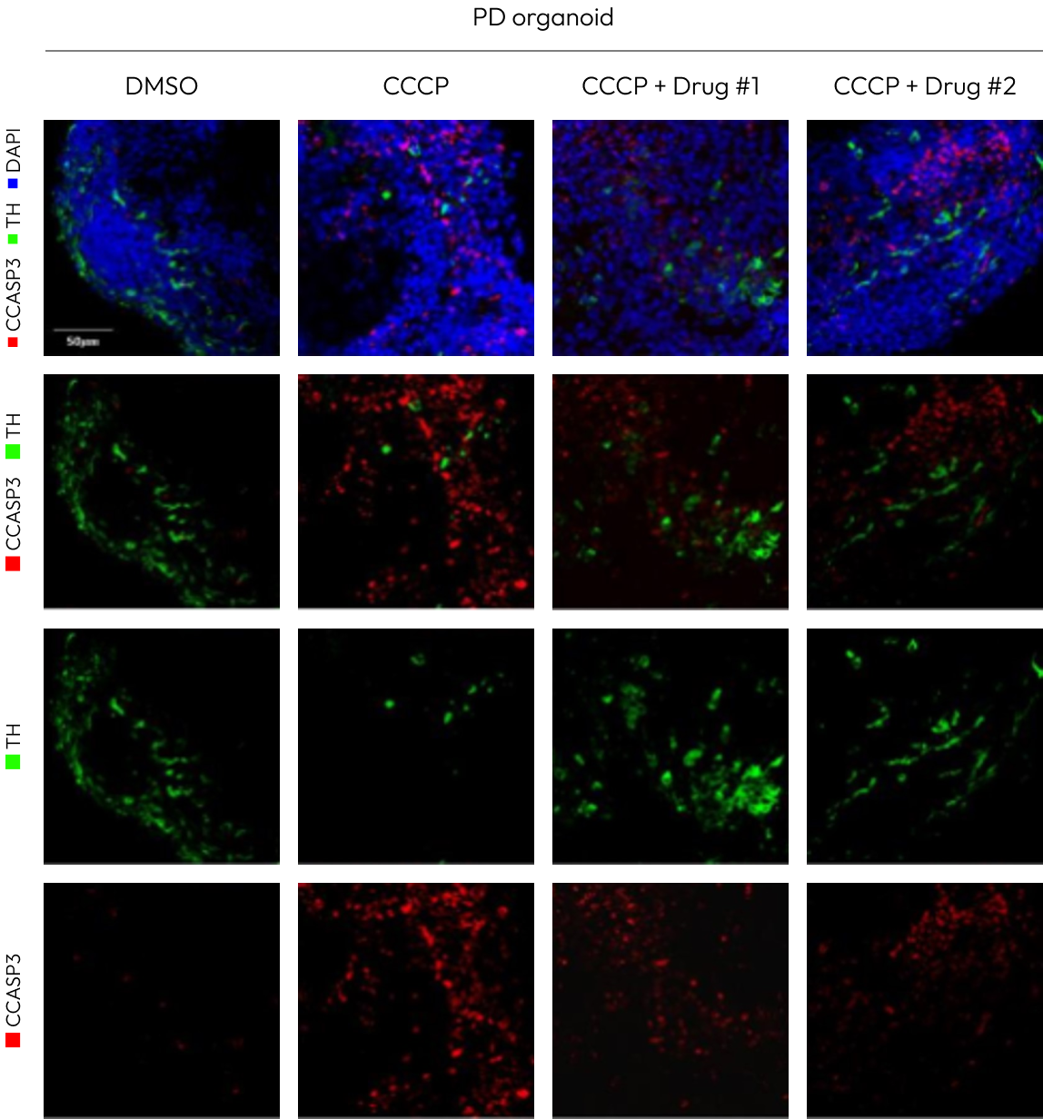
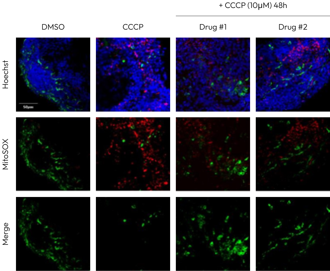
The experiment comparing the expression of th (tyrosine hydroxylase) and ccasp3 (cleaved caspase-3) after treatment with CCCP (a mitochondrial toxin) in Parkinson’s disease organoids is crucial for studying mitochondrial dysfunction and cell death mechanisms.
CCCP depolarizes the mitochondrial membrane potential, inhibiting mitochondrial function and inducing damage to dopamine-producing neurons. In the experiment, th serves as a marker for dopamine-producing neurons, while ccasp3 is a marker for apoptosis.
The results showed a decrease in th expression and an increase in ccasp3 expression following CCCP treatment. This suggests that mitochondrial dysfunction induced by CCCP leads to damage to dopamine neurons and enhances cell death. These findings help confirm the link between mitochondrial dysfunction and cell death pathways in Parkinson’s disease and may offer valuable insights for developing new therapeutic approaches.

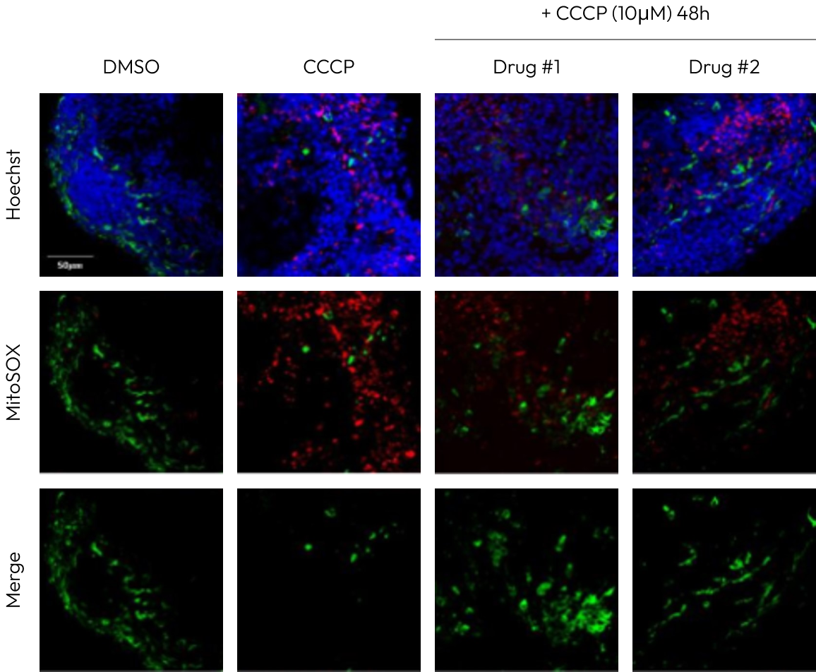
MitoSOX (ROS indicator)

This compound selectively binds to superoxide, a major marker of oxidative stress produced in the mitochondria. After CCCP treatment, the expression of MitoSOX was increased, indicating an elevation in mitochondrial oxidative stress. This supports the notion that mitochondrial dysfunction plays a critical role in Parkinson’s disease.

JC-1 (Mitochondrial membrane potential

JC-1 is used to measure mitochondrial membrane potential. When mitochondria are healthy, JC-1 shifts from green to red fluorescence, while a decrease in membrane potential causes a shift from red to green. After CCCP treatment, there was a tendency for JC-1 expression to shift from red to green, indicating a depolarization of the mitochondrial membrane and loss of function.

Connect with Us