Price | 4929€+ |
Organism | Human |
Product Type | Adult lung tissue-derived organoid, iPSC-derived organoid |
Tissue | Lung |
Disease | – |
Applications
Toxicity
Organoid Based
Anti-Virus
Influenza Virus
Adenovirus
Disease Modeling
Respiratory and ENT Disease
With the global rise of K-beauty, the cosmetics industry continues to grow steadily. Since the ban on animal testing for cosmetics in Korea in 2017, various alternative testing methods have...
Traditional microscopy methods often require fluorescent labeling to analyze cellular structures, which can be time-consuming and invasive. In contrast, our HT-X1 system allows for high-resolution visualization of cellular morphology without...
Traditional protein analysis has primarily focused on quantifying expression levels within tissue samples. However, recent advances in spatial analysis techniques have shifted attention toward evaluating not only expression levels, but...
Among the many fermented foods we consume, kimchi is particularly known for containing a diverse range of lactic acid bacteria, which are believed to influence the activation of immune cells...
We conducted a study focused on identifying disease-related markers using patient-derived tissue samples. However, traditional methods limited our ability to analyze multiple candidate markers simultaneously, and the limited availability of...
Tissue-derived lung organoids are cultured from cells directly obtained from patient lung tissue or biopsy samples, allowing them to closely mimic the cellular composition and microenvironment of the original lung tissue. Because they preserve the genetic expression profile and cell-to-cell interactions of the native tissue, they excel at replicating individual patient physiology. This high degree of similarity to human lungs makes them invaluable for personalized therapy development and disease modeling, offering superior physiological relevance compared to traditional 2D cell cultures or animal models.
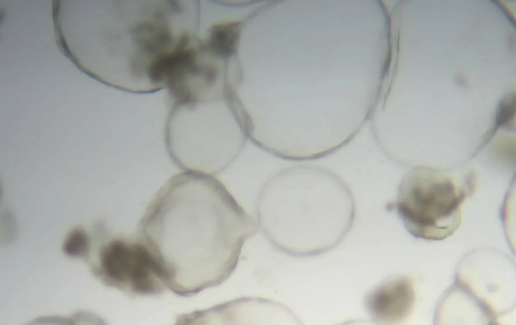
Our lung organoids closely mimic the key structural features of human lung tissue.
Replicating the morphology of actual lung tissue, including critical structures like alveoli.
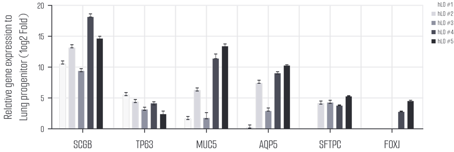
Composed of various cell types, including type I and II alveolar epithelial cells, ciliated cells, and secretory cells, accurately reflecting the functional complexity of real lung tissue.
This compound selectively binds to superoxide, a major marker of oxidative stress produced in the mitochondria. After CCCP treatment, the expression of MitoSOX was increased, indicating an elevation in mitochondrial oxidative stress. This supports the notion that mitochondrial dysfunction plays a critical role in Parkinson’s disease.
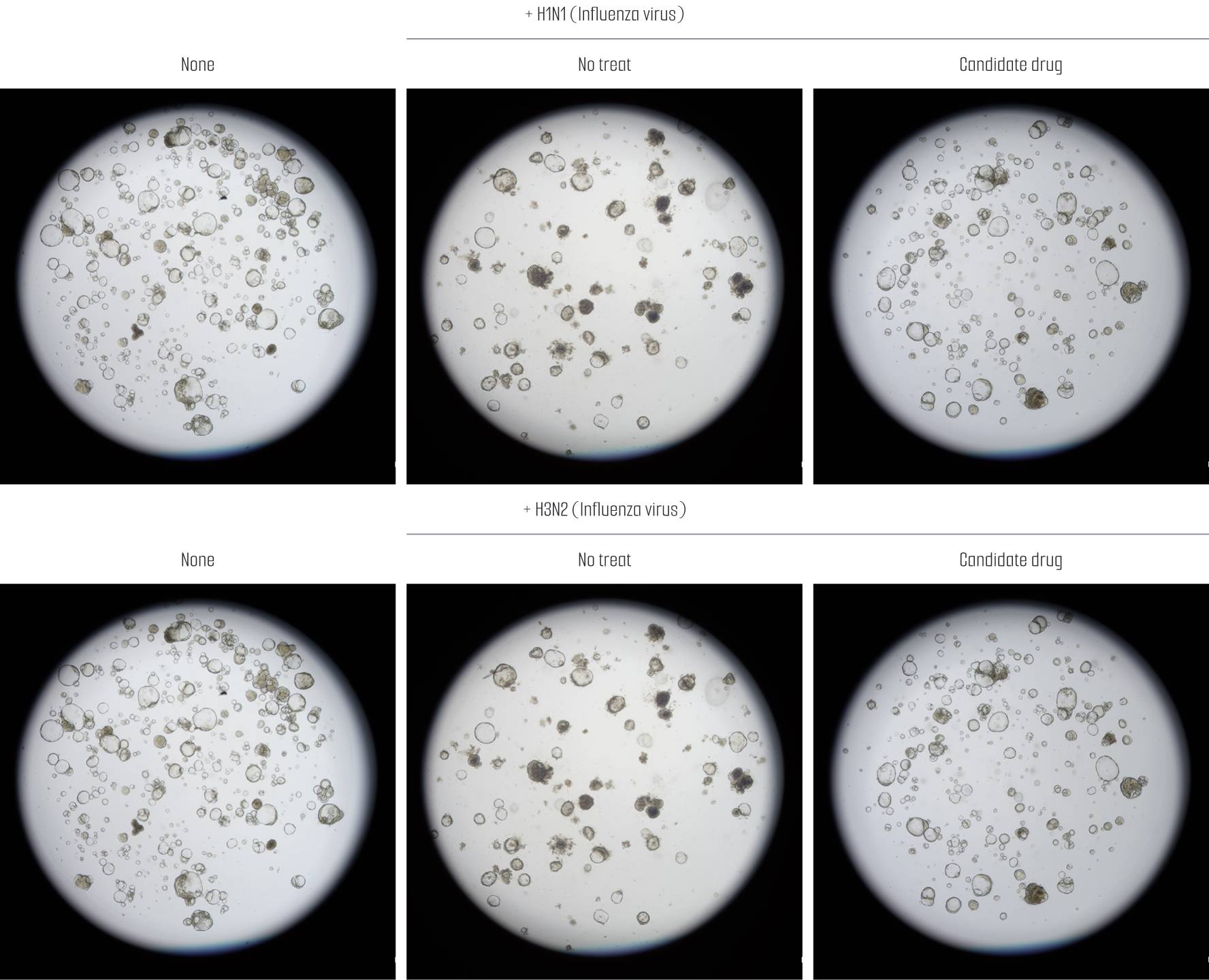
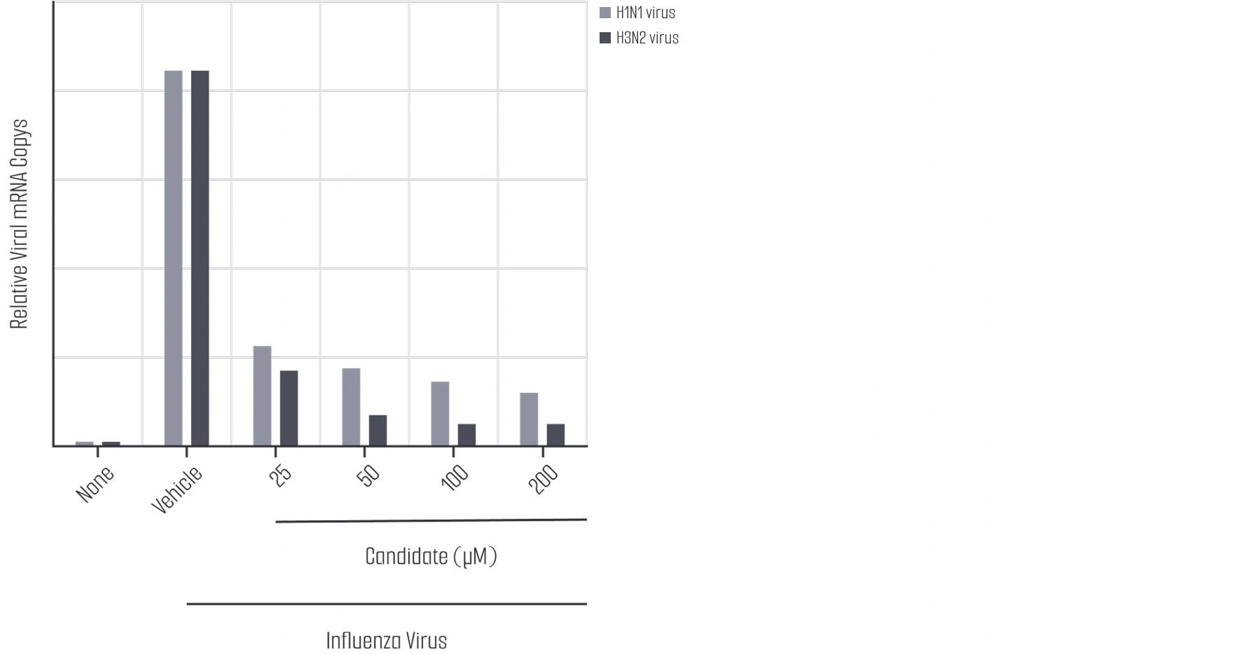
After infecting PSC-derived lung organoids with H1N1 and H3N2 influenza viruses and treating them with candidate drugs at different concentrations (25, 50, 100, 200 µM), RT-PCR analysis revealed a dose-dependent reduction in viral RNA copy numbers. Notably, a significant decrease in viral RNA copy numbers was observed in the candidate drug-treated groups for both H1N1 and H3N2 influenza, suggesting that these candidate drugs may effectively inhibit influenza virus replication.
In the tissue-derived lung organoids with fibrosis induced by TGF-β, immunohistochemical analysis revealed that Nintedanib treatment led to a reduction in the expression of fibrosis markers, A-SMA and Vimentin, confirming its antifibrotic effect. Similar trends were observed in some candidate drug groups, with certain candidates showing a significant decrease in fibrosis marker expression, suggesting potential antifibrotic effects. These findings indicate that Nintedanib and specific candidate drugs may have potential as therapeutic agents for pulmonary fibrosis.
Lambda Biologics GmbH
Deutscher Platz 5 c, 04103, Leipzig, Germany
info@lambdabiologics.com