Search
Lacrimal Gland Organoid_main
Normal Organoid

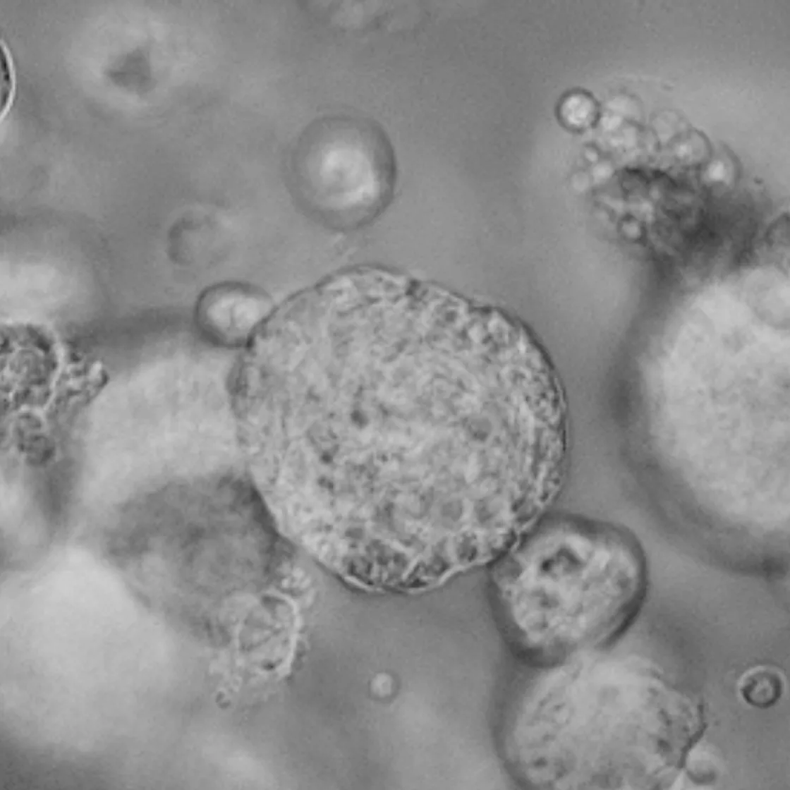
Lacrimal Gland Organoid

4850€+
Normal Organoid

Lacrimal Gland Organoid

  • Histologically similar to lacrimal gland tissue
  • Expression of lacrimal gland-specific markers is similar to that in normal lacrimal gland tissue.
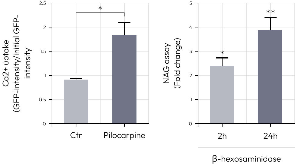
  • Functional response to pilocarpine with increased internal Ca2+ and β-hexosaminidase levels.

Price
Organism
Human
Product Type
Organoid
Tissue
Normal lacrimal gland tissues
Disease

Applications

Toxicity

Organoid Based

Disease Modeling

Gland Disease Model

Description

Table of Contents

Comprehensive Staining Reveals Key Features in Lacrimal Gland Organoids

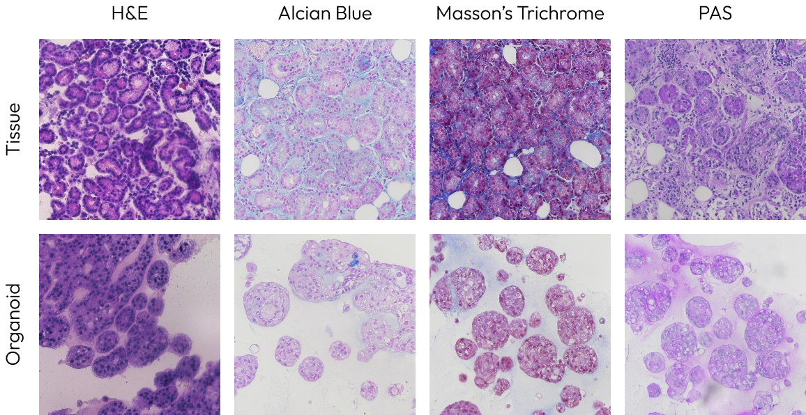
In the lacrimal gland organoid, the structure of acinar cells and their secretion of acidic mucins were observed by H&E, Alcian blue.
Additionally, through PAS staining, secretions such as glycogen and glycoproteins were observed in the organoid.
Masson’s Trichrome staining demonstrated that keratin is a major ECM component in the organoid.

Histology Data

Acinar Cell-Like Expression Patterns of Key Markers in Organoids

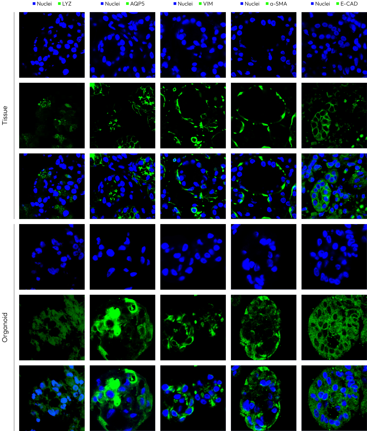
The expression patterns of VIM, AQP5, and LYZ within the organoid closely resemble those of the tissue’s acinar cells.
Furthermore, the expression patterns of E-CAD and α-SMA in the organoid are similar to those in the tissue, though at lower levels.
These markers are not expressed in ductal cells but are expressed in acinar cells, confirming that the formed organoid closely resembles acinar cells.

IF Data

Assessment of Secretory Function in Lacrimal Gland Organoids with Pilocarpine Treatment

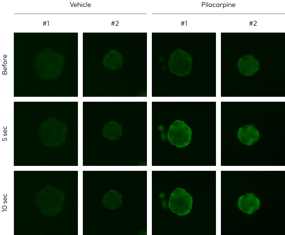
To assess the secretory function, lacrimal gland organoids were treated with pilocarpine.
The intracellular concentration of Ca ions increased, confirming the elevation of the lysosomal protein β-hexosaminidase, a component of tears.

Connect with Us

By submitting your details, you confirm that you have reviewed and agree with the Lambda Biologics Privacy Policy.