Price | 880€+ Login to see price |
Organism | Human |
Product Type | iPS-derived organoid, Adult Tissue derived organoid |
Tissue | Colon, Small Intestine |
Disease | IBD, Inflammatory Bowel Disease, Leaky Gut Syndrome, Infectious enteric disease, Intestinal fibrosis model |
Applications
-
I have been utilizing tumor organoid technology since 2020. Recently, I’ve come to appreciate how combining organoid technology with AI can revolutionize the drug discovery process by enhancing efficiency and...
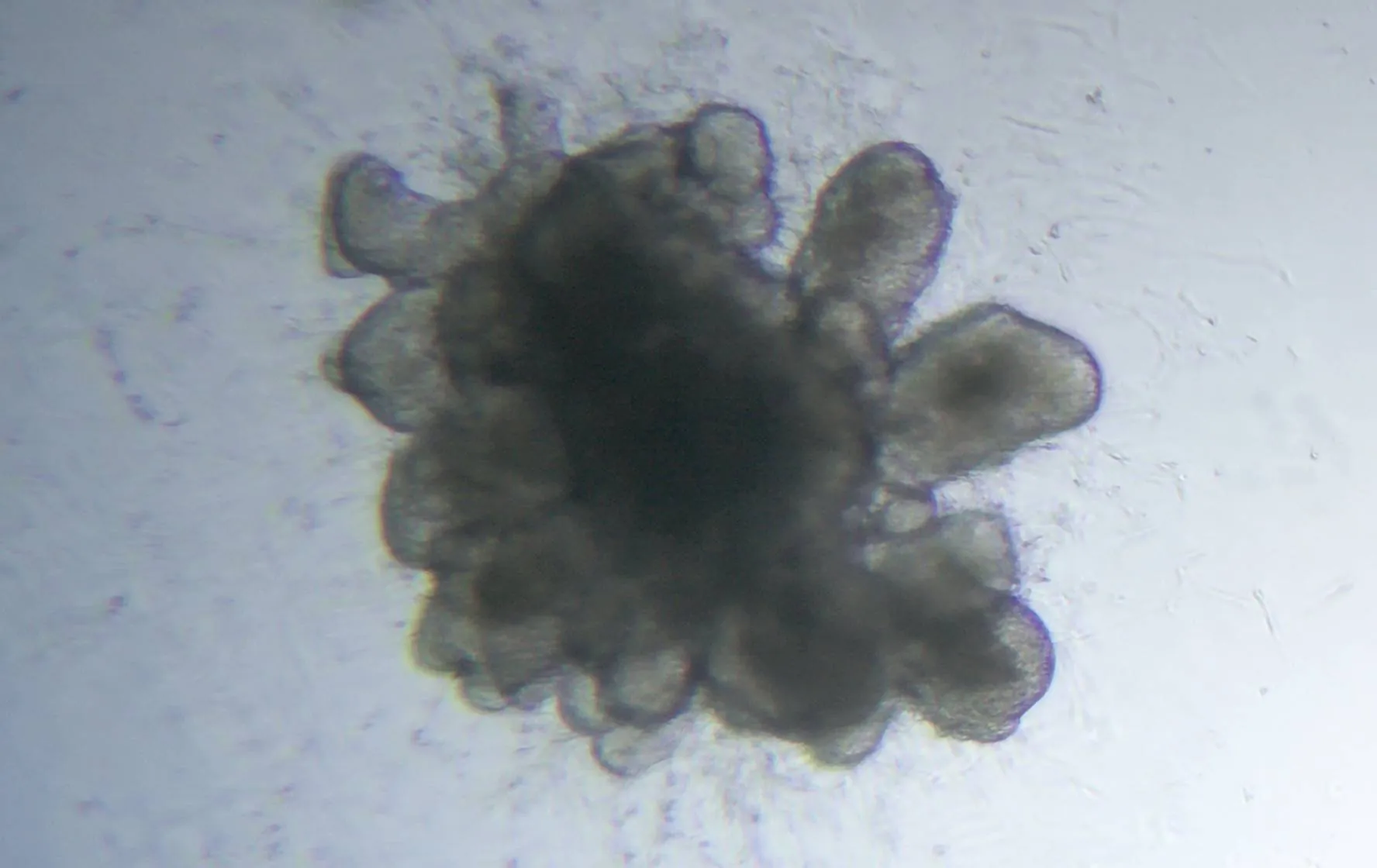
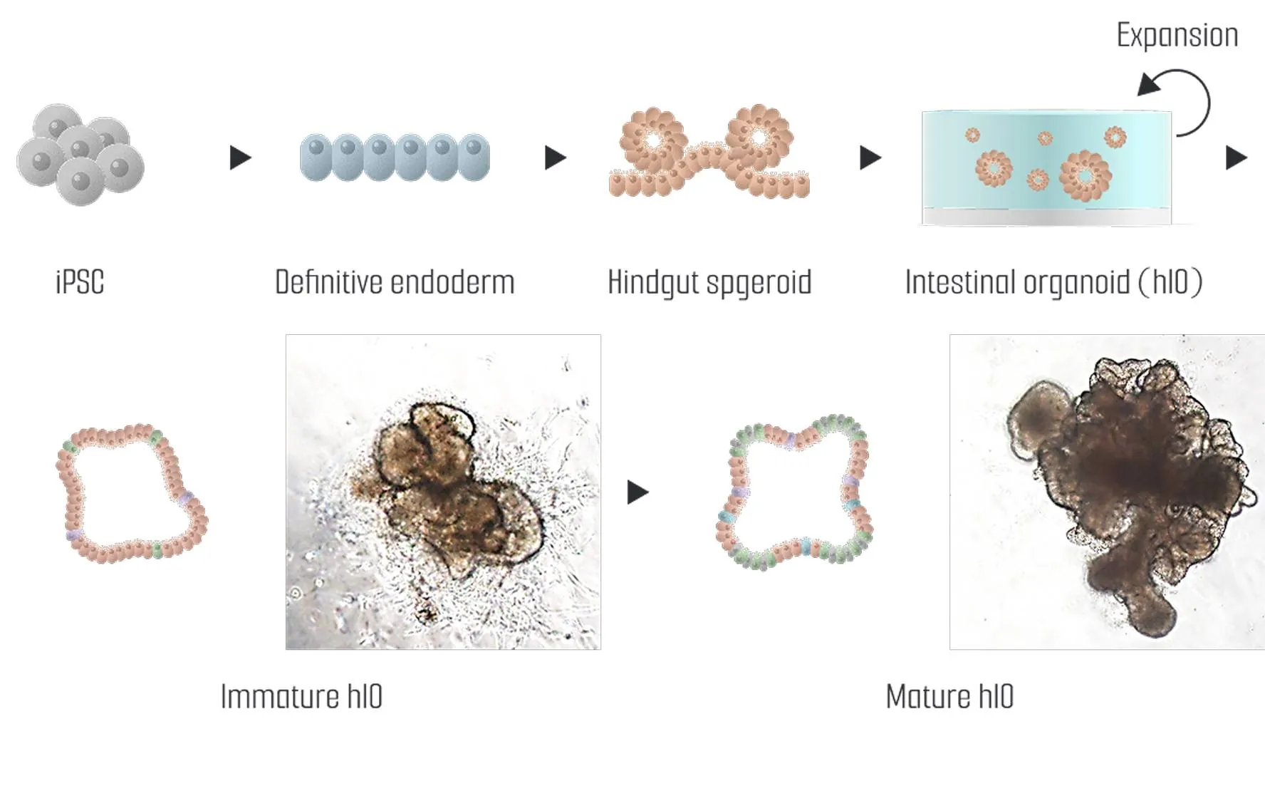
iPSC-derived intestinal organoids are 3D models that replicate intestinal tissue using induced pluripotent stem cells (iPSCs), playing a key role in intestinal development and disease research. These organoids are generated by differentiating iPSCs into intestinal progenitor cells and cultivating them in a suitable extracellular matrix to form 3D structures. The organoids mature into structures with key intestinal layers and functions, which can be assessed through their absorption capacity, bacterial adhesion ability, and drug responses, mimicking real intestinal tissue.
Tissue-derived intestinal organoids are 3D models created using intestinal cells directly isolated from tissue, providing a valuable tool for studying intestinal development, function, and diseases. These organoids are typically generated by culturing intestinal epithelial cells derived from intestinal tissue. Under specialized culture conditions, the cells form 3D structures that closely mimic the natural architecture and function of the intestine. By providing extracellular matrix (ECM) components and growth factors, the cells spontaneously organize into the key layers and morphology of the intestine, maintaining a microenvironment similar to that of the native tissue. These organoids are widely used for modeling intestinal diseases, drug testing, and studying both physiological and pathological conditions of the intestine.
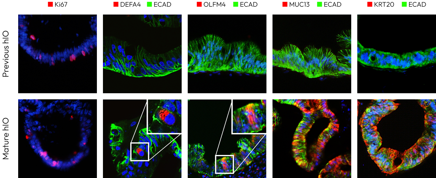
The small intestine is a crucial organ responsible for nutrient absorption, consisting of four layers: the mucosa, submucosa, muscularis propria, and serosa. Inside the small intestine, villi maximize nutrient absorption, while crypts serve as regions where intestinal epithelial cells proliferate. Additionally, microvilli further expand the surface area for absorption. The major cell types include enterocytes responsible for nutrient uptake, goblet cells that secrete mucus, Paneth cells that produce antimicrobial peptides, enteroendocrine cells that release hormones, and intestinal stem cells that regenerate the intestinal epithelium. Intestinal organoids have a structure and cellular composition similar to human intestinal tissue and mimic the functions of the small intestine.
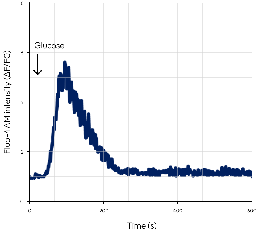
The calcium imaging analysis showed that upon glucose stimulation, mature hIOs exhibited a rapid increase in ΔF/F₀ values, forming a peak followed by a gradual decline. In contrast, previous hIOs displayed weak calcium signaling with consistently low ΔF/F₀ values. This indicates that glucose sensing and signaling pathways in enteroendocrine cells are functionally active in mature hIOs.
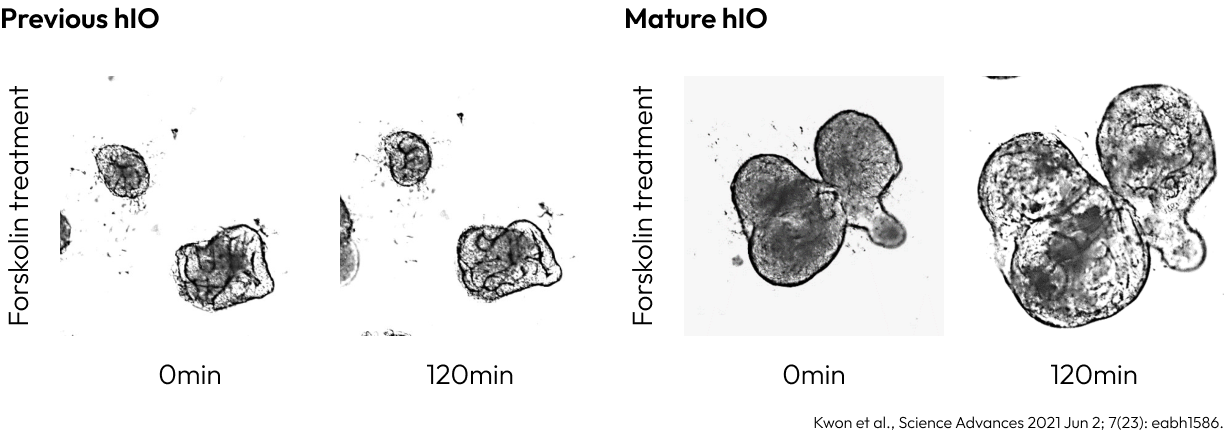
Previous hIO
Mature hIO
The swelling assay revealed that upon forskolin treatment, mature hIOs displayed significant volumetric changes over 120 minutes, while previous hIOs showed minimal or no swelling. This demonstrates that ion transport and water movement via CFTR are active in mature hIOs, effectively replicating the physiological properties of intestinal epithelial cells.
Deutscher Platz 5c, 04103, Leipzig, Germany
info@lambdabiologics.com