Search
Holotomography

Cytotoxicity

1000€+
Holotomography

Cytotoxicity

  • Cell Viability and Death Studies
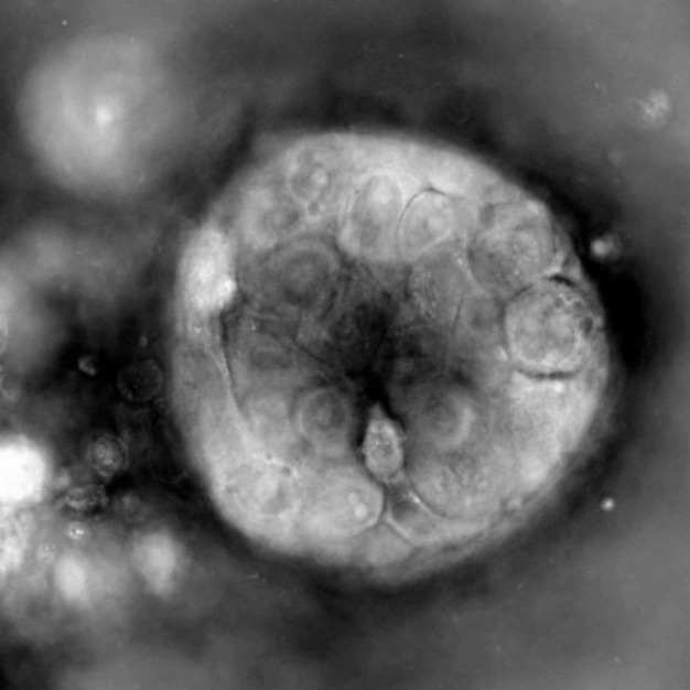
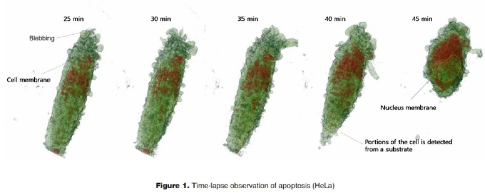
    HT technology allows for 3D observation of cellular morphology without the need for labeling. This is useful for cytotoxicity studies, enabling quantitative analysis of structural changes and cell death processes.
  • Non-invasive 3D Imaging
    HT provides 3D visualization of cells without labeling or staining, allowing for the measurement of refractive index, which can be used to quantify protein concentration, dry mass, and more.
  • Neuronal Cells and Parkinson’s Disease Research
    HT can be used to analyze morphological and physiological changes in neuronal cells in a Parkinson’s disease model, allowing for quantitative classification of cell states.
  • Monitoring of Cell Death Induced by Photosensitizers
    HT allows the monitoring of cell death processes induced by photosensitizers, observing changes such as cell shrinkage, membrane protrusion, and stress granule formation.
  • Versatile Experimental Design
    HT can be used to design experiments for monitoring cell morphology and proliferation, observing early responses to chemical treatments, quantitatively assessing cell volume, dry mass, concentration, and sphericity, 3D observation of organelle positioning and morphological changes, and time-course observation of cytosolic contraction and relaxation movements.

Price
1000€+
Organism
Product Type
Tissue
Disease

Applications

Holotomography

Cell biology

Professor Lee Chang-seok Eulji University
Customer insight

Advancing K-Beauty with Skin Organoids: A Next-Generation Platform for Non-Animal Testing and High-Precision Cosmetic Innovation

With the global rise of K-beauty, the cosmetics industry continues to grow steadily. Since the ban on animal testing for cosmetics in Korea in 2017, various alternative testing methods have...

Tomocube (Spatial)
Customer insight

HT-X1: A Label-Free Imaging Breakthrough for Organoid-Based Disease Modeling and Drug Screening

Traditional microscopy methods often require fluorescent labeling to analyze cellular structures, which can be time-consuming and invasive. In contrast, our HT-X1 system allows for high-resolution visualization of cellular morphology without...

Seoul National University College of Medicine
Customer insight

Pioneering Spatial Protein Analysis in Korea: Advancing Clinical Pathology with Lambda Biologics’ Support

Traditional protein analysis has primarily focused on quantifying expression levels within tissue samples. However, recent advances in spatial analysis techniques have shifted attention toward evaluating not only expression levels, but...

K Research Institute
Customer insight

ODISEI-Gut Platform Reveals Immune-Boosting Potential of Kimchi-Derived Bacterial Strain

Among the many fermented foods we consume, kimchi is particularly known for containing a diverse range of lactic acid bacteria, which are believed to influence the activation of immune cells...

Bundang Jesaeng General Hospital
Customer insight

Multiplex Marker Analysis Enhances Research Efficiency with 31-Marker Detection on a Single Slide

We conducted a study focused on identifying disease-related markers using patient-derived tissue samples. However, traditional methods limited our ability to analyze multiple candidate markers simultaneously, and the limited availability of...

Description

Table of Contents

Cytotoxicity

HT microscopes, using 3D holotomography, allow for label-free observation of individual cells over time, making them ideal for studying cell physiology.
This technology is useful for cell viability and toxicity studies, enabling researchers to monitor cell morphology and proliferation, observe early chemical responses, perform quantitative cell analysis, visualize 3D organelle changes, and track cell movement over time.

Read out

Cytotoxicity_01
Cytotoxicity_02

Connect with Us