Price | 2388€+ Login to see price |
Organism | Human |
Product Type | iPS-derived organoid |
Tissue | Brain (Cerebral) |
Disease | Neurodegenerative diseases, Brain tumor, Neurodevelopmental disorders |
Applications
With the global rise of K-beauty, the cosmetics industry continues to grow steadily. Since the ban on animal testing for cosmetics in Korea in 2017, various alternative testing methods have...
Traditional microscopy methods often require fluorescent labeling to analyze cellular structures, which can be time-consuming and invasive. In contrast, our HT-X1 system allows for high-resolution visualization of cellular morphology without...
Traditional protein analysis has primarily focused on quantifying expression levels within tissue samples. However, recent advances in spatial analysis techniques have shifted attention toward evaluating not only expression levels, but...
Among the many fermented foods we consume, kimchi is particularly known for containing a diverse range of lactic acid bacteria, which are believed to influence the activation of immune cells...
We conducted a study focused on identifying disease-related markers using patient-derived tissue samples. However, traditional methods limited our ability to analyze multiple candidate markers simultaneously, and the limited availability of...
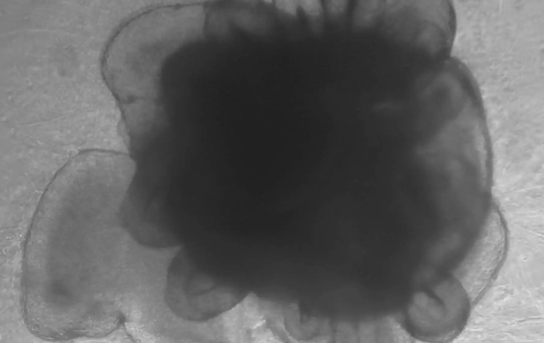
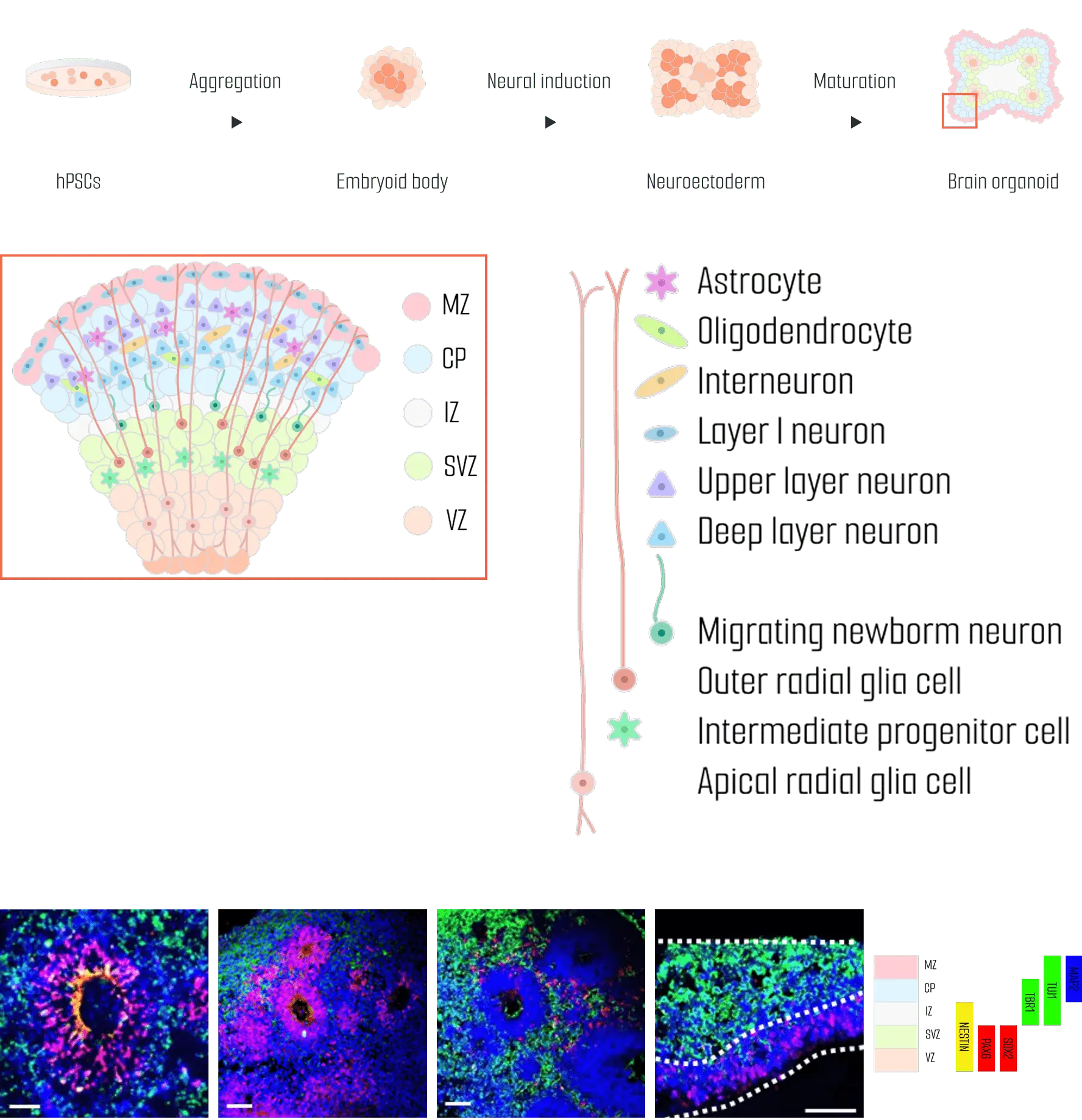
We have developed highly functional organoids capable of long-term culture by applying our maturation technology to human brain organoids derived from hPSCs.
VZ (Ventricular Zone) – Contains neural stem cells and serves as the birthplace of neurons and glial cells.
SVZ (Subventricular Zone) – A region where neural progenitor cells differentiate and prepare for migration.
IZ (Intermediate Zone) – The migratory pathway for neurons, where axons and neural fibers are formed.
CP (Cortical Plate) – The region where mature neurons settle to form the cortical structure.
MZ (Marginal Zone) – The outermost layer of the cerebral cortex involved in neural circuit formation and signal modulation.
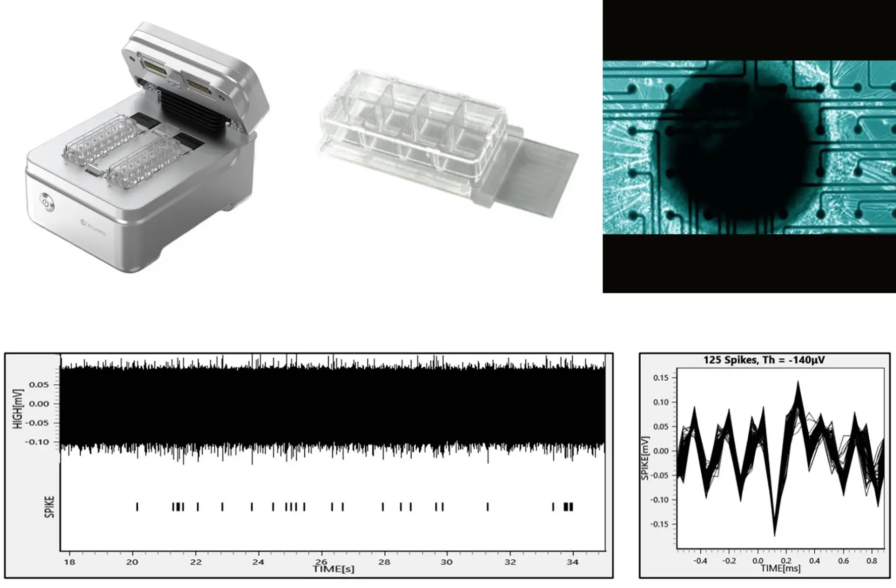
The FP data obtained through electrophysiological analysis provides valuable insights into the neural network activity of the organoid. The FP measured in the natural, untreated state reflects the organoid’s ability to generate electrical activity independently, providing evidence that it has formed a functional neural network. This data is essential for understanding how organoids can be used in neuroscience research, disease modeling, and neural network studies.
Mature brain organoids exhibit well-formed and connected neural networks, showing consistent electrical activity. The field potentials (FP) generated by the organoid are measured through the electrode array on the chip, capturing the electrical activity produced by the interactions between neural cells. These voltage changes occur when multiple neural cells are simultaneously activated. In mature organoids, it is typically observed that firing occurs naturally at a frequency of 0.1–1 Hz, reflecting the rhythmic activity of the neural network.
We can generate organoid models of neurodegenerative diseases through three methods: creating brain disease organoids using iPSCs with regulated expression of specific genes or inducing brain disease organoids through drug treatment.
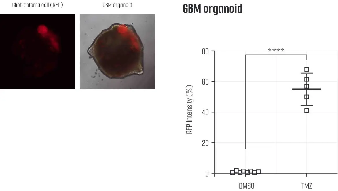
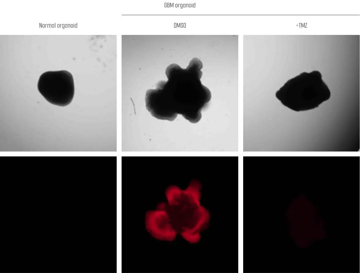
Glioblastoma multiforme (GBM) is the most aggressive form of brain tumor, characterized by rapid growth and resistance to treatment. It primarily occurs in the cerebral hemispheres and severely affects brain function. GBM tumors have poorly defined borders, exhibit active angiogenesis, and display a complex mixture of cell types. The main treatments include surgery, radiation therapy, and chemotherapy, but recurrence rates are high, and prognosis is generally poor.
Deutscher Platz 5c, 04103, Leipzig, Germany
info@lambdabiologics.com