Fibrosis is a disease characterized by repetitive injury and inflammation.
Lambda provides a lung and intestine fibrosis organoid model, enabling drug development and efficacy assessment.


Fibrosis
Lambda’s Intestinal Fibrosis Model addresses these challenges by leveraging high mimetic human intestinal organoids to create a model of inflammatory bowel diseases. This model facilitates the evaluation of drug penetration concentrations and efficacy. It proves particularly valuable in the development of anti-fibrotic drugs, providing a reliable platform for assessing their efficacy.
Cell Type
· iPSC
Organoid type
· Intestinal organoid
Lambda’s IPF organoid model provides a highly mimetic representation of the structural and functional characteristics of human lung tissue, serving as a crucial platform for studying the mechanisms of pulmonary fibrosis, testing drug efficacy, and researching personalized treatment approaches.
Cell Type
· ASC
Organoid type
· Lung organoid

Idiopathic pulmonary fibrosis(IPF) is a chronic lung disease characterized by progressive scarring of lung tissue for which the cause is unknown.
In IPF, fibrotic tissue accumulates in the lungs, gradually impairing their function.
Lambda’s IPF organoid model provides a highly mimetic representation of the structural and functional characteristics of human lung tissue, serving as a crucial platform for studying the mechanisms of pulmonary fibrosis, testing drug efficacy, and researching personalized treatment approaches.


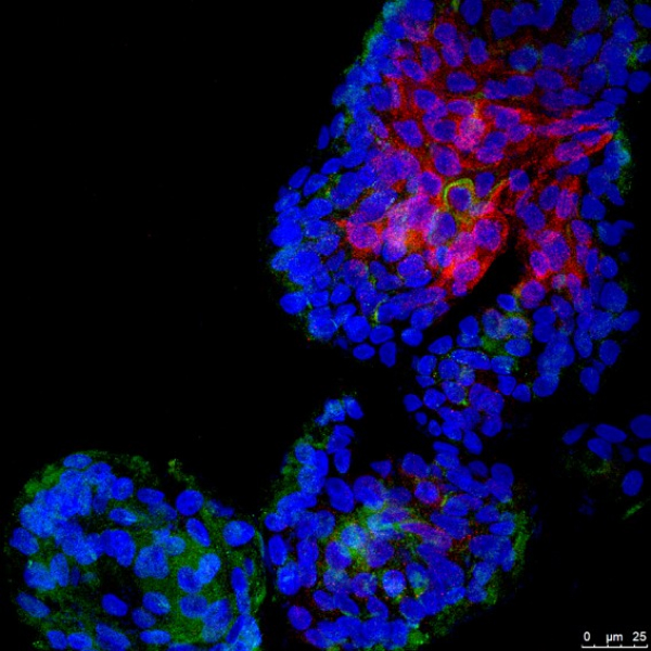
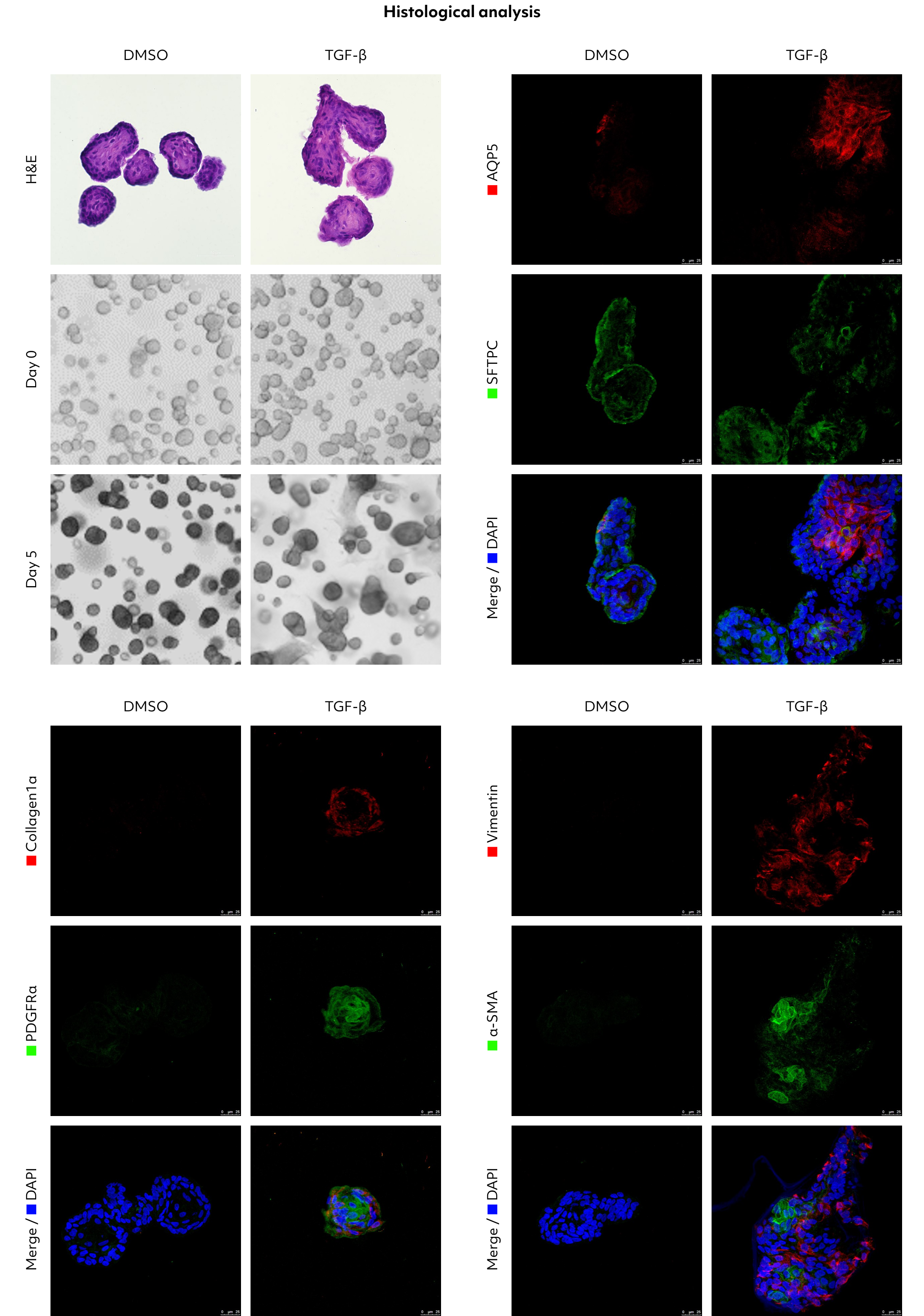
Compare the morphological characteristics of the control group and the fibrosis group using H&E staining.
Furthermore, assess the presence of TGF-beta, a substance associated with fibrosis,
in both the control and fibrosis groups using Immunofluorescence (IF) analysis.

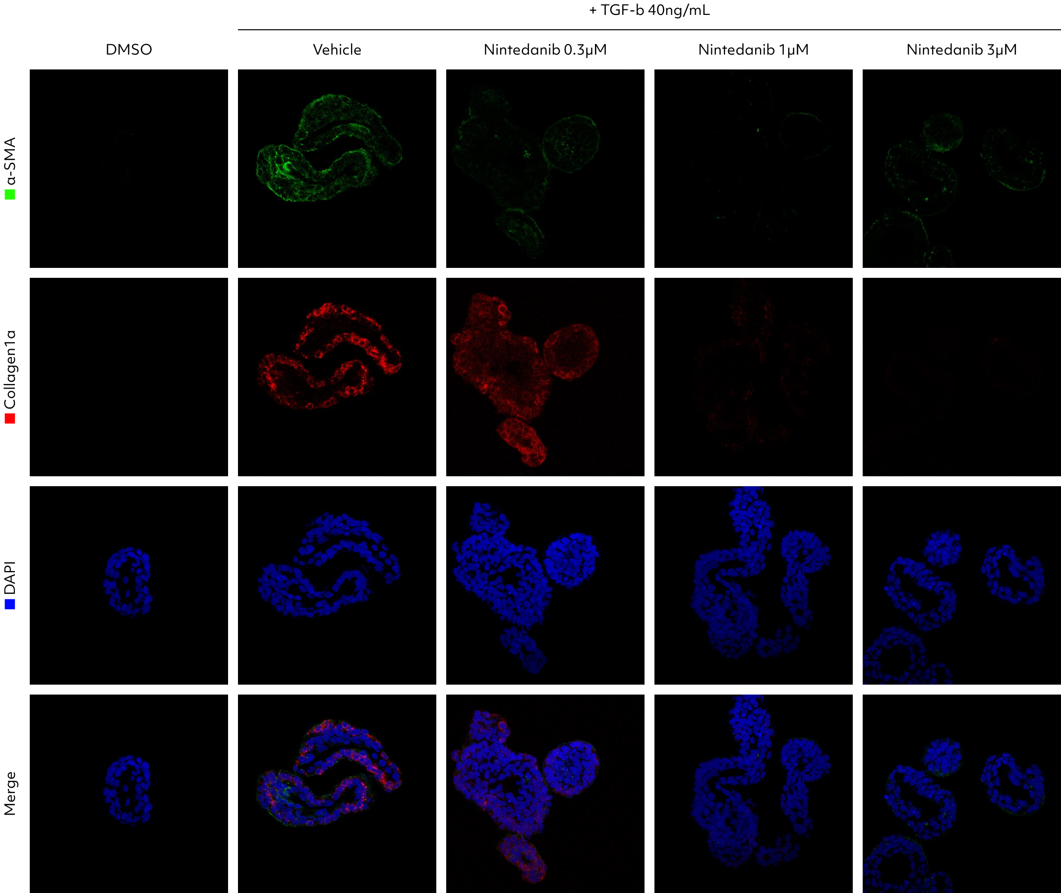
In the fibrosis model, we verified the reduction of the TGF-beta effect by treating it with Nintedanib, a recognized inhibitor of TGF-beta.
This confirmation was achieved through the analysis of collagen type 1a and ɑ-SMA markers, using Immunofluorescence (IF).