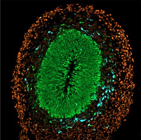
Within the elegant convergence of art and scientific inquiry, cerebral organoids emerge as revolutionary tools transforming our understanding of neurological development. Alex Kingston’s award-nominated photograph from Imperial College London’s competition captures this breakthrough technology with striking visual clarity — showcasing a single rosette within a cerebral organoid through fluorescent antibody staining.
These laboratory-cultivated “mini-brains” represent a significant advancement in biotechnology. Each organoid contains dozens of these intricate rosettes, functioning as microscopic models of human brain development in its earliest stages. The vibrant green identifies progenitor cells while orange marks neuronal identity, creating a biological tapestry that reveals fundamental developmental processes.
Kingston’s research focuses on how cells engineered to disrupt environmental sensing (shown in blue) behave within complex tissues. This investigation into physical forces during development opens pathways to understanding neurological disorders and potential therapeutic interventions, exemplifying how visualisation techniques and advanced biological models are propelling biotechnology toward increasingly precise and impactful discoveries